Movie
Movie Controller

[English] 日本語
 Yorodumi
- EMDB-45846: Focused map of PAXX/XLF in a gap-filling complex with Pol mu enga... -
+ Open data

- Basic information
| Entry |  | |||||||||
|---|---|---|---|---|---|---|---|---|---|---|
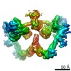
| Title | Focused map of PAXX/XLF in a gap-filling complex with Pol mu engaged in the NHEJ pathway | |||||||||
 Map data Map data | focused PAXX map in a gap-filling complex in the NHEJ pathway | |||||||||
 Sample Sample |
| |||||||||
 Keywords Keywords | NHEJ / DNA gap / fill-in synthesis / ligation / XLF / PAXX / Polymerase mu / DNA repair / Ligase IV / DNA BINDING PROTEIN | |||||||||
| Biological species |  Homo sapiens (human) Homo sapiens (human) | |||||||||
| Method | single particle reconstruction / cryo EM / Resolution: 7.4 Å | |||||||||
 Authors Authors | Li J / Liu L / Gellert M / Yang W | |||||||||
| Funding support |  United States, 1 items United States, 1 items
| |||||||||
 Citation Citation | Journal: Nature / Year: 2025 Title: Dynamic assemblies and coordinated reactions of non-homologous end joining. Authors: Lan Liu / Jun Li / Metztli Cisneros-Aguirre / Arianna Merkell / Jeremy M Stark / Martin Gellert / Wei Yang / Abstract: Non-homologous end joining (NHEJ) is the main repair pathway of double-strand DNA breaks in higher eukaryotes. Here we report reconstitution of the final steps of NHEJ and structures of DNA ...Non-homologous end joining (NHEJ) is the main repair pathway of double-strand DNA breaks in higher eukaryotes. Here we report reconstitution of the final steps of NHEJ and structures of DNA polymerase μ and ligase IV (LIG4) engaged in gap filling and end joining. These reactions take place in a flexible ω-shaped framework composed of XRCC4 and XLF. Two broken DNA ends, each encircled by Ku70-Ku80 internally, are docked onto the ω frame, mediated by LIG4. DNA polymerase and ligase attached to each ω arm repair only one broken strand of a defined polarity; the final steps of NHEJ requires coordination and toggling of a pair of such enzymes. The facilitators XLF and PAXX additively stimulate NHEJ reactions. As DNA-end sensor and protector, LIG4 replaces DNA-PKcs for end joining and bridges the two DNA ends for polymerase to fill remaining gaps. These assemblies present new targets for NHEJ inhibition to enhance efficacy of radiotherapy and accuracy of gene editing. | |||||||||
| History |
|
- Structure visualization
| Supplemental images |
|---|

- Downloads & links
-EMDB archive
| Map data | emd_45846.map.gz | 407.4 MB |  EMDB map data format EMDB map data format | |
|---|---|---|---|---|
| Header (meta data) | emd-45846-v30.xmlemd-45846.xml | 17.9 KB 17.9 KB | Display Display | EMDB header |
| FSC (resolution estimation) | emd_45846_fsc.xml | 18.4 KB | Display | FSC data file |
| Images |  emd_45846.png emd_45846.png | 42.6 KB | ||
| Masks | emd_45846_msk_1.map | 512 MB | Mask map | |
| Filedesc metadata | emd-45846.cif.gz | 5.3 KB | ||
| Others | emd_45846_half_map_1.map.gzemd_45846_half_map_2.map.gz | 410.8 MB 410.8 MB | ||
| Archive directory |  http://ftp.pdbj.org/pub/emdb/structures/EMD-45846ftp://ftp.pdbj.org/pub/emdb/structures/EMD-45846 http://ftp.pdbj.org/pub/emdb/structures/EMD-45846ftp://ftp.pdbj.org/pub/emdb/structures/EMD-45846 | HTTPS FTP |
-Validation report
| Summary document | emd_45846_validation.pdf.gz | 847.2 KB | Display | EMDB validaton report |
|---|---|---|---|---|
| Full document | emd_45846_full_validation.pdf.gz | 846.7 KB | Display | |
| Data in XML | emd_45846_validation.xml.gz | 25.5 KB | Display | |
| Data in CIF | emd_45846_validation.cif.gz | 34.3 KB | Display | |
| Arichive directory | https://ftp.pdbj.org/pub/emdb/validation_reports/EMD-45846ftp://ftp.pdbj.org/pub/emdb/validation_reports/EMD-45846 | HTTPS FTP |
-Related structure data


-Links
| EMDB pages | EMDB (EBI/PDBe) / EMDataResource |
|---|
-Map
| File | Download / File: emd_45846.map.gz / Format: CCP4 / Size: 512 MB / Type: IMAGE STORED AS FLOATING POINT NUMBER (4 BYTES) | ||||||||||||||||||||||||||||||||||||
|---|---|---|---|---|---|---|---|---|---|---|---|---|---|---|---|---|---|---|---|---|---|---|---|---|---|---|---|---|---|---|---|---|---|---|---|---|---|
| Annotation | focused PAXX map in a gap-filling complex in the NHEJ pathway | ||||||||||||||||||||||||||||||||||||
| Projections & slices | Image control
Images are generated by Spider. | ||||||||||||||||||||||||||||||||||||
| Voxel size | X=Y=Z: 0.833 Å | ||||||||||||||||||||||||||||||||||||
| Density |
| ||||||||||||||||||||||||||||||||||||
| Symmetry | Space group: 1 | ||||||||||||||||||||||||||||||||||||
| Details | EMDB XML:
|
 Z (Sec.)
 Y (Row.)
 X (Col.)


-Supplemental data
-Mask #1
| File | emd_45846_msk_1.map | ||||||||||||
|---|---|---|---|---|---|---|---|---|---|---|---|---|---|
| Projections & Slices |
| ||||||||||||
| Density Histograms |


-Half map: half 1 map
| File | emd_45846_half_map_1.map | ||||||||||||
|---|---|---|---|---|---|---|---|---|---|---|---|---|---|
| Annotation | half 1 map | ||||||||||||
| Projections & Slices |
| ||||||||||||
| Density Histograms |


-Half map: half 2 map
| File | emd_45846_half_map_2.map | ||||||||||||
|---|---|---|---|---|---|---|---|---|---|---|---|---|---|
| Annotation | half 2 map | ||||||||||||
| Projections & Slices |
| ||||||||||||
| Density Histograms |


- Sample components
-Entire : A gap-filling complex with Pol mu engaged
| Entire | Name: A gap-filling complex with Pol mu engaged |
|---|---|
| Components |
|
-Supramolecule #1: A gap-filling complex with Pol mu engaged
| Supramolecule | Name: A gap-filling complex with Pol mu engaged / type: complex / ID: 1 / Parent: 0 / Macromolecule list: all |
|---|---|
| Source (natural) | Organism: Homo sapiens (human) |
| Molecular weight | Theoretical: 967 KDa |
-Macromolecule #1: human PAXX
| Macromolecule | Name: human PAXX / type: protein_or_peptide / ID: 1 / Enantiomer: LEVO |
|---|---|
| Source (natural) | Organism: Homo sapiens (human) |
| Recombinant expression | Organism:  |
| Sequence | String: MGSSHHHHHH SQDPMDPLSP PLCTLPPGPE PPRFVCYCEG EESGEGDRGG FNLYVTDAAE LWSTCFTPDS LAAL KARFG LSAAEDITPR FRAACEQQAV ALTLQEDRAS LTLSGGPSAL AFDLSKVPGP EAAPR LRAL TLGLAKRVWS LERRLAAAEE TAVSPRKSPR ...String: MGSSHHHHHH SQDPMDPLSP PLCTLPPGPE PPRFVCYCEG EESGEGDRGG FNLYVTDAAE LWSTCFTPDS LAAL KARFG LSAAEDITPR FRAACEQQAV ALTLQEDRAS LTLSGGPSAL AFDLSKVPGP EAAPR LRAL TLGLAKRVWS LERRLAAAEE TAVSPRKSPR PAGPQLFLPD PDPQRGGPGP GVRRRC PGE SLINPGFKSK KPAGGVDFDE T |
-Macromolecule #2: human XLF
| Macromolecule | Name: human XLF / type: protein_or_peptide / ID: 2 / Enantiomer: LEVO |
|---|---|
| Source (natural) | Organism: Homo sapiens (human) |
| Recombinant expression | Organism: Homo sapiens (human) |
| Sequence | String: GPVMEELEQG LLMQPWAWLQ LAENSLLAKV FITKQGYALL VSDLQQVWHE QVDTSVVSQR AKE LNKRLT APPAAFLCHL DNLLRPLLKD AAHPSEATFS CDCVADALIL RVRSELSGLP FYWN FHCML ASPSLVSQHL IRPLMGMSLA LQCQVRELAT LLHMKDLEIQ ...String: GPVMEELEQG LLMQPWAWLQ LAENSLLAKV FITKQGYALL VSDLQQVWHE QVDTSVVSQR AKE LNKRLT APPAAFLCHL DNLLRPLLKD AAHPSEATFS CDCVADALIL RVRSELSGLP FYWN FHCML ASPSLVSQHL IRPLMGMSLA LQCQVRELAT LLHMKDLEIQ DYQESGATLI RDRLK TEPF EENSFLEQFM IEKLPEACSI GDGKPFVMNL QDLYMAVTTQ EVQVGQKHQG AGDPHT SNS ASLQGIDSQC VNQPEQLVSS APTLSAPEKE STGTSGPLQR PQLSKVKRKK PRGLFS |
-Experimental details
-Structure determination
| Method | cryo EM |
|---|---|
 Processing Processing | single particle reconstruction |
| Aggregation state | particle |
-Sample preparation
| Concentration | 0.35 mg/mL |
|---|---|
| Buffer | pH: 7.9 |
| Vitrification | Cryogen name: ETHANE / Chamber humidity: 100 % / Chamber temperature: 277 K |
- Electron microscopy
| Microscope | FEI TITAN KRIOS |
|---|---|
| Image recording | Film or detector model: GATAN K3 BIOQUANTUM (6k x 4k) / Average electron dose: 54.4 e/Å2 |
| Electron beam | Acceleration voltage: 300 kV / Electron source:  FIELD EMISSION GUN FIELD EMISSION GUN |
| Electron optics | Illumination mode: FLOOD BEAM / Imaging mode: BRIGHT FIELD / Nominal defocus max: 1.5 µm / Nominal defocus min: 0.5 µm |
| Experimental equipment |  Model: Titan Krios / Image courtesy: FEI Company |