 Movie
Movie Controller

[English] 日本語
 Yorodumi
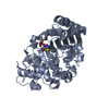
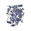
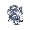
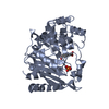
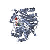
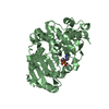
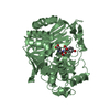
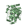
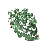
- PDB-1llb: Crystal Structure Of AmpC beta-Lactamase From E. Coli In Complex ... -
+ Open data

- Basic information
| Entry | Database: PDB / ID: 1llb | ||||||
|---|---|---|---|---|---|---|---|
| Title | Crystal Structure Of AmpC beta-Lactamase From E. Coli In Complex With ATMO-penicillin | ||||||
 Components Components | beta-lactamase | ||||||
 Keywords Keywords | HYDROLASE / cephalosporinase / beta-lactamase / serine | ||||||
| Function / homology |  Function and homology information Function and homology informationantibiotic catabolic process / beta-lactamase activity / beta-lactamase / outer membrane-bounded periplasmic space / response to antibiotic Similarity search - Function | ||||||
| Biological species |  | ||||||
| Method |  X-RAY DIFFRACTION / SYNCHROTRON / MOLECULAR REPLACEMENT / Resolution: 1.72 Å X-RAY DIFFRACTION / SYNCHROTRON / MOLECULAR REPLACEMENT / Resolution: 1.72 Å | ||||||
 Authors Authors | Trehan, I. / Morandi, F. / Blaszczak, L.C. / Shoichet, B.K. | ||||||
 Citation Citation | Journal: Chem.Biol. / Year: 2002 Title: Using steric hindrance to design new inhibitors of class C beta-lactamases. Authors: Trehan, I. / Morandi, F. / Blaszczak, L.C. / Shoichet, B.K. | ||||||
| History |
| ||||||
| Remark 600 | heterogen The het group PCN was origianlly ATMO-penicillin or 6-[Z-(2-aminothiazol-4-yl)- ...heterogen The het group PCN was origianlly ATMO-penicillin or 6-[Z-(2-aminothiazol-4-yl)-methoximinoacetamido]- penicillanic acid before the compound underwent a nucleophilic attack at the C1 by the Ser64 residue and is now covalently bound to Ser64. This attack breaks the bond between C1 and N7, thus opening the 4-membered ring. |
- Structure visualization
| Structure viewer | Molecule: MolmilJmol/JSmol |
|---|
- Downloads & links
-Download
| PDBx/mmCIF format | 1llb.cif.gz | 164 KB | Display | PDBx/mmCIF format |
|---|---|---|---|---|
| PDB format | pdb1llb.ent.gz | 128.5 KB | Display | PDB format |
| PDBx/mmJSON format | 1llb.json.gz | Tree view | PDBx/mmJSON format | |
| Others |  Other downloads Other downloads |
-Validation report
| Arichive directory | https://data.pdbj.org/pub/pdb/validation_reports/ll/1llbftp://data.pdbj.org/pub/pdb/validation_reports/ll/1llb | HTTPS FTP |
|---|
-Related structure data
| Related structure data | 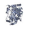 1ll9C 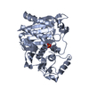 1ke4S S: Starting model for refinement C: citing same article ( |
|---|---|
| Similar structure data |


-Links
 PDBj
PDBj
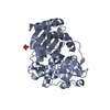
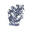
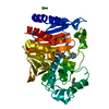
- Assembly
| Deposited unit | 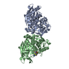
| ||||||||
|---|---|---|---|---|---|---|---|---|---|
| 1 | 
| ||||||||
| 2 | 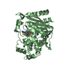
| ||||||||
| Unit cell |
|
-Components
| #1: Protein | Mass: 39587.922 Da / Num. of mol.: 2 Source method: isolated from a genetically manipulated source Source: (gene. exp.) #2: Chemical | ChemComp-PCN / |   Mass: 401.461 Da / Num. of mol.: 1 / Source method: obtained synthetically / Formula: C14H19N5O5S2 Mass: 401.461 Da / Num. of mol.: 1 / Source method: obtained synthetically / Formula: C14H19N5O5S2#3: Water | ChemComp-HOH / |  Mass: 18.015 Da / Num. of mol.: 598 / Source method: isolated from a natural source / Formula: H2O Mass: 18.015 Da / Num. of mol.: 598 / Source method: isolated from a natural source / Formula: H2OHas protein modification | Y | |
|---|
-Experimental details
-Experiment
| Experiment | Method: X-RAY DIFFRACTION / Number of used crystals: 1 |
|---|
- Sample preparation
| Crystal | Density Matthews: 2.53 Å3/Da / Density % sol: 51.4 % |
|---|---|
| Crystal grow | Temperature: 296 K / Method: vapor diffusion, hanging drop / pH: 8.7 Details: potassium phosphate, AmpC, pH 8.7, VAPOR DIFFUSION, HANGING DROP, temperature 296K |
| Crystal grow | *PLUS Temperature: 23 ℃ / Details: Trehan, I., (2001) Biochemistry, 40, 7992. |
| Components of the solutions | *PLUS Conc.: 1.7 M / Common name: potassium phosphate |
-Data collection
| Diffraction | Mean temperature: 100 K |
|---|---|
| Diffraction source | Source: SYNCHROTRON / Site: APS  / Beamline: 5ID-B / Wavelength: 1 Å / Beamline: 5ID-B / Wavelength: 1 Å |
| Detector | Type: MARRESEARCH / Detector: CCD / Date: Jun 7, 2001 |
| Radiation | Protocol: SINGLE WAVELENGTH / Monochromatic (M) / Laue (L): M / Scattering type: x-ray |
| Radiation wavelength | Wavelength: 1 Å / Relative weight: 1 |
| Reflection | Resolution: 1.72→20 Å / Num. all: 81383 / Num. obs: 81383 / % possible obs: 97 % / Observed criterion σ(I): -3 / Rmerge(I) obs: 0.035 / Net I/σ(I): 31.49 |
| Reflection shell | Resolution: 1.72→1.76 Å / Rmerge(I) obs: 0.243 / Mean I/σ(I) obs: 4.36 / % possible all: 95.3 |
| Reflection | *PLUS Lowest resolution: 20 Å / % possible obs: 97 % / Num. measured all: 270758 / Rmerge(I) obs: 0.035 |
| Reflection shell | *PLUS % possible obs: 95.3 % / Rmerge(I) obs: 0.243 |
- Processing
| Software |
| |||||||||||||||||||||||||
|---|---|---|---|---|---|---|---|---|---|---|---|---|---|---|---|---|---|---|---|---|---|---|---|---|---|---|
| Refinement | Method to determine structure: MOLECULAR REPLACEMENT Starting model: PDB entry 1KE4 with solvent atoms removed Resolution: 1.72→20 Å / σ(F): 0 / σ(I): 2 / Stereochemistry target values: Engh & Huber
| |||||||||||||||||||||||||
| Refinement step | Cycle: LAST / Resolution: 1.72→20 Å
| |||||||||||||||||||||||||
| Refine LS restraints |
| |||||||||||||||||||||||||
| Refinement | *PLUS Lowest resolution: 20 Å / Rfactor Rfree: 0.199 / Rfactor Rwork: 0.178 | |||||||||||||||||||||||||
| Solvent computation | *PLUS | |||||||||||||||||||||||||
| Displacement parameters | *PLUS | |||||||||||||||||||||||||
| Refine LS restraints | *PLUS
|
