Movie
Movie Controller

[English] 日本語
 Yorodumi
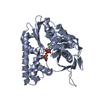
- EMDB-1470: Molecular structure of the ParM polymer and the mechanism leading... -
+ Open data

- Basic information
| Entry | Database: EMDB / ID: EMD-1470 | |||||||||
|---|---|---|---|---|---|---|---|---|---|---|
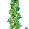
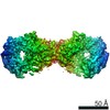
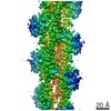
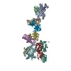
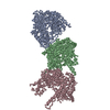
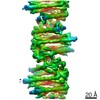
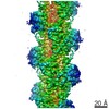
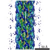
| Title | Molecular structure of the ParM polymer and the mechanism leading to its nucleotide-driven dynamic instability | |||||||||
 Map data Map data | A volume file of ParM filament | |||||||||
 Sample Sample |
| |||||||||
 Keywords Keywords | GTPase / molecular switch / filament / ParM | |||||||||
| Function / homology | Plasmid segregation protein ParM/StbA / : / : / Plasmid segregation protein ParM, N-terminal / Plasmid segregation protein ParM, C-terminal / plasmid partitioning / ATPase, nucleotide binding domain / identical protein binding / Plasmid segregation protein ParM Function and homology information Function and homology information | |||||||||
| Biological species |  | |||||||||
| Method | helical reconstruction / negative staining / Resolution: 23.0 Å | |||||||||
 Authors Authors | Popp D / Narita A / Oda T / Fujisawa T / Matsuo H / Nitanai Y / Iwasa M / Maeda K / Onishi H / Maeda Y | |||||||||
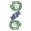
 Citation Citation | Journal: EMBO J / Year: 2008 Title: Molecular structure of the ParM polymer and the mechanism leading to its nucleotide-driven dynamic instability. Authors: David Popp / Akihiro Narita / Toshiro Oda / Tetsuro Fujisawa / Hiroshi Matsuo / Yasushi Nitanai / Mitsusada Iwasa / Kayo Maeda / Hirofumi Onishi / Yuichiro Maéda /  Abstract: ParM is a prokaryotic actin homologue, which ensures even plasmid segregation before bacterial cell division. In vivo, ParM forms a labile filament bundle that is reminiscent of the more complex ...ParM is a prokaryotic actin homologue, which ensures even plasmid segregation before bacterial cell division. In vivo, ParM forms a labile filament bundle that is reminiscent of the more complex spindle formed by microtubules partitioning chromosomes in eukaryotic cells. However, little is known about the underlying structural mechanism of DNA segregation by ParM filaments and the accompanying dynamic instability. Our biochemical, TIRF microscopy and high-pressure SAX observations indicate that polymerization and disintegration of ParM filaments is driven by GTP rather than ATP and that ParM acts as a GTP-driven molecular switch similar to a G protein. Image analysis of electron micrographs reveals that the ParM filament is a left-handed helix, opposed to the right-handed actin polymer. Nevertheless, the intersubunit contacts are similar to those of actin. Our atomic model of the ParM-GMPPNP filament, which also fits well to X-ray fibre diffraction patterns from oriented gels, can explain why after nucleotide release, large conformational changes of the protomer lead to a breakage of intra- and interstrand interactions, and thus to the observed disintegration of the ParM filament after DNA segregation. | |||||||||
| History |
|
- Structure visualization
| Movie |
Movie viewer |
|---|---|
| Structure viewer | EM map: SurfViewMolmilJmol/JSmol |
| Supplemental images |
 UCSF Chimera
- Downloads & links
-EMDB archive
| Map data | emd_1470.map.gz | 196.2 KB | EMDB map data format | |
|---|---|---|---|---|
| Header (meta data) | emd-1470-v30.xmlemd-1470.xml | 9.3 KB 9.3 KB | Display Display | EMDB header |
| Images | 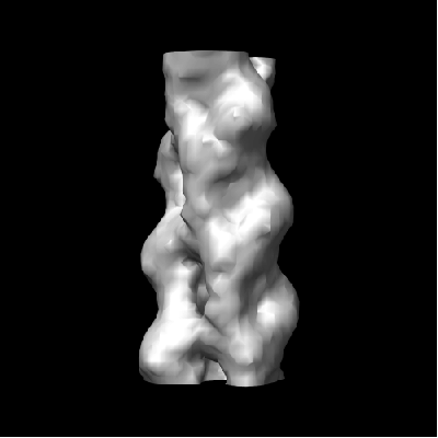 1470.gif 1470.gif | 29.6 KB | ||
| Archive directory |  http://ftp.pdbj.org/pub/emdb/structures/EMD-1470ftp://ftp.pdbj.org/pub/emdb/structures/EMD-1470 http://ftp.pdbj.org/pub/emdb/structures/EMD-1470ftp://ftp.pdbj.org/pub/emdb/structures/EMD-1470 | HTTPS FTP |
-Validation report
| Summary document | emd_1470_validation.pdf.gz | 292.8 KB | Display | EMDB validaton report |
|---|---|---|---|---|
| Full document | emd_1470_full_validation.pdf.gz | 292.4 KB | Display | |
| Data in XML | emd_1470_validation.xml.gz | 3.9 KB | Display | |
| Arichive directory | https://ftp.pdbj.org/pub/emdb/validation_reports/EMD-1470ftp://ftp.pdbj.org/pub/emdb/validation_reports/EMD-1470 | HTTPS FTP |
-Related structure data
| Related structure data | 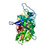 2zhcMC 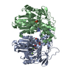 2zgyC 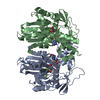 2zgzC M: atomic model generated by this map C: citing same article ( |
|---|---|
| Similar structure data |
-Links
| EMDB pages | EMDB (EBI/PDBe) / EMDataResource |
|---|
-Map
| File | Download / File: emd_1470.map.gz / Format: CCP4 / Size: 257.8 KB / Type: IMAGE STORED AS FLOATING POINT NUMBER (4 BYTES) | ||||||||||||||||||||||||||||||||||||||||||||||||||||||||||||||||||||
|---|---|---|---|---|---|---|---|---|---|---|---|---|---|---|---|---|---|---|---|---|---|---|---|---|---|---|---|---|---|---|---|---|---|---|---|---|---|---|---|---|---|---|---|---|---|---|---|---|---|---|---|---|---|---|---|---|---|---|---|---|---|---|---|---|---|---|---|---|---|
| Annotation | A volume file of ParM filament | ||||||||||||||||||||||||||||||||||||||||||||||||||||||||||||||||||||
| Projections & slices | Image control
Images are generated by Spider. generated in cubic-lattice coordinate | ||||||||||||||||||||||||||||||||||||||||||||||||||||||||||||||||||||
| Voxel size | X=Y=Z: 4.011 Å | ||||||||||||||||||||||||||||||||||||||||||||||||||||||||||||||||||||
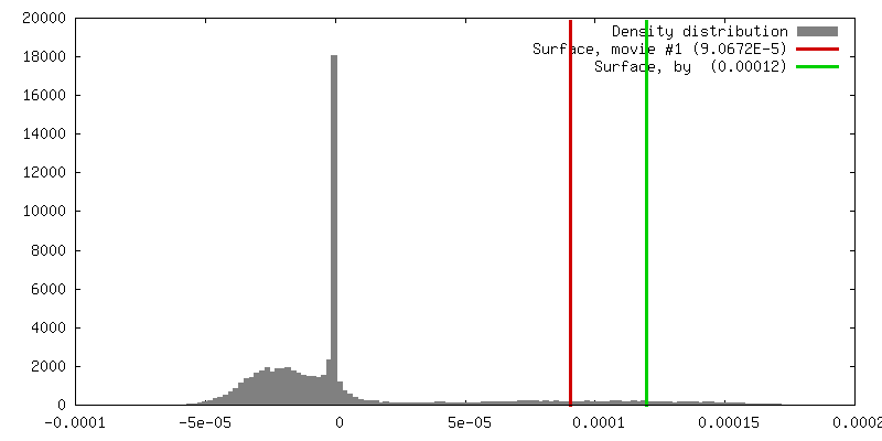
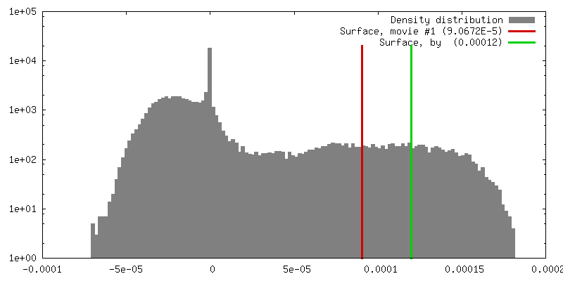
| Density |
| ||||||||||||||||||||||||||||||||||||||||||||||||||||||||||||||||||||
| Symmetry | Space group: 1 | ||||||||||||||||||||||||||||||||||||||||||||||||||||||||||||||||||||
| Details | EMDB XML:
CCP4 map header:
| ||||||||||||||||||||||||||||||||||||||||||||||||||||||||||||||||||||
 Z (Sec.)
 Y (Row.)
 X (Col.)


-Supplemental data
- Sample components
-Entire : ParM filament
| Entire | Name: ParM filament |
|---|---|
| Components |
|
-Supramolecule #1000: ParM filament
| Supramolecule | Name: ParM filament / type: sample / ID: 1000 / Oligomeric state: Filament / Number unique components: 1 |
|---|---|
| Molecular weight | Theoretical: 35 KDa |
-Macromolecule #1: ParM
| Macromolecule | Name: ParM / type: protein_or_peptide / ID: 1 / Name.synonym: ParM / Number of copies: 10 / Oligomeric state: Helical filament / Recombinant expression: Yes |
|---|---|
| Source (natural) | Organism: |
| Recombinant expression | Organism: |
-Experimental details
-Structure determination
| Method | negative staining |
|---|---|
 Processing Processing | helical reconstruction |
| Aggregation state | filament |
-Sample preparation
| Concentration | 0.14 mg/mL |
|---|---|
| Buffer | pH: 7.5 Details: 10 mM Hepes, 25 mM KCl, 1 mM MgCl2, 1 mM DTT, 5 mM GMPPNP |
| Staining | Type: NEGATIVE / Details: 1.0 % uranyl acetate |
| Vitrification | Cryogen name: NONE / Instrument: OTHER |
- Electron microscopy
| Microscope | JEOL 2010HC |
|---|---|
| Image recording | Digitization - Sampling interval: 7 µm / Number real images: 7 |
| Electron beam | Acceleration voltage: 100 kV / Electron source: LAB6 |
| Electron optics | Illumination mode: FLOOD BEAM / Imaging mode: BRIGHT FIELD |
| Sample stage | Specimen holder model: OTHER |
-Image processing
| Final reconstruction | Resolution.type: BY AUTHOR / Resolution: 23.0 Å / Resolution method: FSC 0.5 CUT-OFF / Software - Name: EOS |
|---|---|
| CTF correction | Details: Each filament |