- EMDB-5352: Structure of a type III secretion needle -
+
Open data
ID or keywords:
Loading...
-
Basic information
Entry
Database: EMDB / ID: EMD-5352
Title
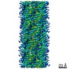
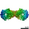
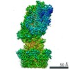
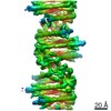
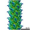
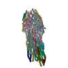
Structure of a type III secretion needle
Map data
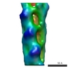
This is an reconstruction of the type III secretion needle
Sample
Sample: Shigella needle
Protein or peptide: MxiH
Keywords
type III secretion system / needle / helical filament
Function / homology
Function and homology information
type III protein secretion system complex / protein secretion by the type III secretion system / cell surface / extracellular region / identical protein binding Similarity search - Function
Type III secretion, needle-protein-like / Type III secretion, needle-protein-like superfamily / Type III secretion needle MxiH, YscF, SsaG, EprI, PscF, EscF / Type III secretion system, needle protein Similarity search - Domain/homology
Type 3 secretion system needle filament protein / Type 3 secretion system needle filament protein Similarity search - Component
Biological species
Shigella flexneri (bacteria)
Method
helical reconstruction / cryo EM / Resolution: 7.7 Å
Journal: Proc Natl Acad Sci U S A / Year: 2012 Title: Structure of a type III secretion needle at 7-Å resolution provides insights into its assembly and signaling mechanisms. Authors: Takashi Fujii / Martin Cheung / Amandine Blanco / Takayuki Kato / Ariel J Blocker / Keiichi Namba / Abstract: Type III secretion systems of Gram-negative bacteria form injection devices that deliver effector proteins into eukaryotic cells during infection. They span both bacterial membranes and the ...Type III secretion systems of Gram-negative bacteria form injection devices that deliver effector proteins into eukaryotic cells during infection. They span both bacterial membranes and the extracellular space to connect with the host cell plasma membrane. Their extracellular portion is a needle-like, hollow tube that serves as a secretion conduit for effector proteins. The needle of Shigella flexneri is approximately 50-nm long and 7-nm thick and is made by the helical assembly of one protein, MxiH. We provide a 7-Å resolution 3D image reconstruction of the Shigella needle by electron cryomicroscopy, which resolves α-helices and a β-hairpin that has never been observed in the crystal and solution structures of needle proteins, including MxiH. An atomic model of the needle based on the 3D-density map, in comparison with that of the bacterial-flagellar filament, provides insights into how such a thin tubular structure is stably assembled by intricate intermolecular interactions. The map also illuminates how the needle-length control protein functions as a ruler within the central channel during export of MxiH for assembly at the distal end of the needle, and how the secretion-activation signal may be transduced through a conformational change of the needle upon host-cell contact.
History
Deposition
Nov 2, 2011
-
Header (metadata) release
Nov 28, 2011
-
Map release
Feb 20, 2012
-
Update
Mar 6, 2012
-
Current status
Mar 6, 2012
Processing site: RCSB / Status: Released
-
Structure visualization
Movie
Surface view with section colored by density value
pH: 7.4 / Details: 20 mM Tris, pH 7.4, 150 mM NaCl, 2mM MgSO4
Vitrification
Cryogen name: ETHANE / Chamber humidity: 100 % / Instrument: OTHER / Details: Vitrification instrument: Vitrobot / Method: Blot for 3.5 seconds before plunging
-
Electron microscopy
Microscope
JEOL 3200FSC
Temperature
Min: 50 K / Max: 60 K / Average: 50 K
Specialist optics
Energy filter - Name: JEOL Omega filter / Energy filter - Lower energy threshold: 0.0 eV / Energy filter - Upper energy threshold: 10.0 eV
Date
Jul 2, 2008
Image recording
Category: CCD / Film or detector model: TVIPS TEMCAM-F415 (4k x 4k) / Digitization - Sampling interval: 15 µm / Number real images: 330 / Average electron dose: 20 e/Å2 / Bits/pixel: 16
Electron beam
Acceleration voltage: 200 kV / Electron source: FIELD EMISSION GUN
In the structure databanks used in Yorodumi, some data are registered as the other names, "COVID-19 virus" and "2019-nCoV". Here are the details of the virus and the list of structure data.
Jan 31, 2019. EMDB accession codes are about to change! (news from PDBe EMDB page)
EMDB accession codes are about to change! (news from PDBe EMDB page)
The allocation of 4 digits for EMDB accession codes will soon come to an end. Whilst these codes will remain in use, new EMDB accession codes will include an additional digit and will expand incrementally as the available range of codes is exhausted. The current 4-digit format prefixed with “EMD-” (i.e. EMD-XXXX) will advance to a 5-digit format (i.e. EMD-XXXXX), and so on. It is currently estimated that the 4-digit codes will be depleted around Spring 2019, at which point the 5-digit format will come into force.
The EM Navigator/Yorodumi systems omit the EMD- prefix.
Related info.:Q: What is EMD? / ID/Accession-code notation in Yorodumi/EM Navigator
Yorodumi is a browser for structure data from EMDB, PDB, SASBDB, etc.
This page is also the successor to EM Navigator detail page, and also detail information page/front-end page for Omokage search.
The word "yorodu" (or yorozu) is an old Japanese word meaning "ten thousand". "mi" (miru) is to see.
Related info.:EMDB / PDB / SASBDB / Comparison of 3 databanks / Yorodumi Search / Aug 31, 2016. New EM Navigator & Yorodumi / Yorodumi Papers / Jmol/JSmol / Function and homology information / Changes in new EM Navigator and Yorodumi
 Movie
Movie Controller


 Open data
 Basic information
 Map data
 Sample
 Keywords
 Function and homology information
 Shigella flexneri (bacteria)
 Authors
 Citation
 Structure visualization
 Downloads & links
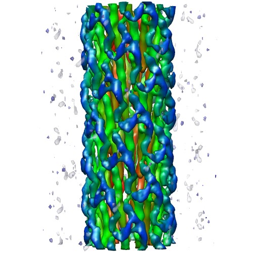 emd_5352_1.jpg
 http://ftp.pdbj.org/pub/emdb/structures/EMD-5352
 Z (Sec.)
 Y (Row.)
 X (Col.)


 Sample components
 Processing
 Electron microscopy
 FIELD EMISSION GUN