 Movie
Movie Controller


+ Open data

- Basic information
| Entry | Database: EMDB / ID: EMD-0397 | |||||||||
|---|---|---|---|---|---|---|---|---|---|---|
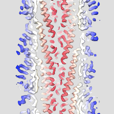
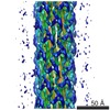
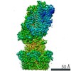
| Title | Cryo-EM reconstruction of Sulfolobus islandicus LAL14/1 Pilus | |||||||||
 Map data Map data | primary map | |||||||||
 Sample Sample |
| |||||||||
 Keywords Keywords | helical symmetry / archaeal pilus / STRUCTURAL PROTEIN | |||||||||
| Function / homology | DUF973 family protein Function and homology information Function and homology information | |||||||||
| Biological species |   Sulfolobus islandicus LAL14/1 (archaea) Sulfolobus islandicus LAL14/1 (archaea) | |||||||||
| Method | helical reconstruction / cryo EM / Resolution: 4.1 Å | |||||||||
 Authors Authors | Wang F / Cvirkaite-Krupovic V | |||||||||
| Funding support |  United States, 1 items United States, 1 items
| |||||||||
 Citation Citation | Journal: Nat Microbiol / Year: 2019 Title: An extensively glycosylated archaeal pilus survives extreme conditions. Authors: Fengbin Wang / Virginija Cvirkaite-Krupovic / Mark A B Kreutzberger / Zhangli Su / Guilherme A P de Oliveira / Tomasz Osinski / Nicholas Sherman / Frank DiMaio / Joseph S Wall / David ...Authors: Fengbin Wang / Virginija Cvirkaite-Krupovic / Mark A B Kreutzberger / Zhangli Su / Guilherme A P de Oliveira / Tomasz Osinski / Nicholas Sherman / Frank DiMaio / Joseph S Wall / David Prangishvili / Mart Krupovic / Edward H Egelman /  Abstract: Pili on the surface of Sulfolobus islandicus are used for many functions, and serve as receptors for certain archaeal viruses. The cells grow optimally at pH 3 and ~80 °C, exposing these ...Pili on the surface of Sulfolobus islandicus are used for many functions, and serve as receptors for certain archaeal viruses. The cells grow optimally at pH 3 and ~80 °C, exposing these extracellular appendages to a very harsh environment. The pili, when removed from cells, resist digestion by trypsin or pepsin, and survive boiling in sodium dodecyl sulfate or 5 M guanidine hydrochloride. We used electron cryo-microscopy to determine the structure of these filaments at 4.1 Å resolution. An atomic model was built by combining the electron density map with bioinformatics without previous knowledge of the pilin sequence-an approach that should prove useful for assemblies where all of the components are not known. The atomic structure of the pilus was unusual, with almost one-third of the residues being either threonine or serine, and with many hydrophobic surface residues. While the map showed extra density consistent with glycosylation for only three residues, mass measurements suggested extensive glycosylation. We propose that this extensive glycosylation renders these filaments soluble and provides the remarkable structural stability. We also show that the overall fold of the archaeal pilin is remarkably similar to that of archaeal flagellin, establishing common evolutionary origins. | |||||||||
| History |
|
- Structure visualization
| Movie |
Movie viewer |
|---|---|
| Structure viewer | EM map: SurfViewMolmilJmol/JSmol |
| Supplemental images |
- Downloads & links
-EMDB archive
| Map data | emd_0397.map.gz | 12 MB | EMDB map data format | |
|---|---|---|---|---|
| Header (meta data) | emd-0397-v30.xmlemd-0397.xml | 10 KB 10 KB | Display Display | EMDB header |
| Images |  emd_0397.png emd_0397.png | 180.3 KB | ||
| Filedesc metadata | emd-0397.cif.gz | 5 KB | ||
| Archive directory |  http://ftp.pdbj.org/pub/emdb/structures/EMD-0397ftp://ftp.pdbj.org/pub/emdb/structures/EMD-0397 http://ftp.pdbj.org/pub/emdb/structures/EMD-0397ftp://ftp.pdbj.org/pub/emdb/structures/EMD-0397 | HTTPS FTP |
-Related structure data
| Related structure data |  6navMC M: atomic model generated by this map C: citing same article ( |
|---|---|
| Similar structure data |
-Links
| EMDB pages | EMDB (EBI/PDBe) / EMDataResource |
|---|
-Map
| File | Download / File: emd_0397.map.gz / Format: CCP4 / Size: 216 MB / Type: IMAGE STORED AS FLOATING POINT NUMBER (4 BYTES) | ||||||||||||||||||||||||||||||||||||||||||||||||||||||||||||
|---|---|---|---|---|---|---|---|---|---|---|---|---|---|---|---|---|---|---|---|---|---|---|---|---|---|---|---|---|---|---|---|---|---|---|---|---|---|---|---|---|---|---|---|---|---|---|---|---|---|---|---|---|---|---|---|---|---|---|---|---|---|
| Annotation | primary map | ||||||||||||||||||||||||||||||||||||||||||||||||||||||||||||
| Projections & slices | Image control
Images are generated by Spider. | ||||||||||||||||||||||||||||||||||||||||||||||||||||||||||||
| Voxel size | X=Y=Z: 1.4 Å | ||||||||||||||||||||||||||||||||||||||||||||||||||||||||||||
| Density |
| ||||||||||||||||||||||||||||||||||||||||||||||||||||||||||||
| Symmetry | Space group: 1 | ||||||||||||||||||||||||||||||||||||||||||||||||||||||||||||
| Details | EMDB XML:
CCP4 map header:
| ||||||||||||||||||||||||||||||||||||||||||||||||||||||||||||
 Z (Sec.)
 Y (Row.)
 X (Col.)


-Supplemental data
- Sample components
-Entire : Sulfolobus islandicus LAL14/1 Pilus
| Entire | Name: Sulfolobus islandicus LAL14/1 Pilus |
|---|---|
| Components |
|
-Supramolecule #1: Sulfolobus islandicus LAL14/1 Pilus
| Supramolecule | Name: Sulfolobus islandicus LAL14/1 Pilus / type: complex / ID: 1 / Parent: 0 / Macromolecule list: all |
|---|---|
| Source (natural) | Organism: Sulfolobus islandicus LAL14/1 (archaea) |
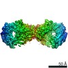
-Macromolecule #1: M9UD72
| Macromolecule | Name: M9UD72 / type: protein_or_peptide / ID: 1 / Number of copies: 21 / Enantiomer: LEVO |
|---|---|
| Source (natural) | Organism: Sulfolobus islandicus LAL14/1 (archaea) |
| Molecular weight | Theoretical: 12.681375 KDa |
| Sequence | String: LSGAVTALIL VIASVIIALV VVGFAFGLFG AFTGQGTVTQ VGTATLSAST GTLKVTLKNT GATTQVTGAI INGNAASVSG QVTISAGQS TYSISLGGIS SSTLQSLVGS TISLTLQLSN GQTVTVSAII TS UniProtKB: DUF973 family protein |
-Experimental details
-Structure determination
| Method | cryo EM |
|---|---|
 Processing Processing | helical reconstruction |
| Aggregation state | filament |
-Sample preparation
| Buffer | pH: 6 |
|---|---|
| Vitrification | Cryogen name: ETHANE / Chamber humidity: 90 % |
- Electron microscopy
| Microscope | FEI TITAN KRIOS |
|---|---|
| Image recording | Film or detector model: FEI FALCON III (4k x 4k) / Detector mode: INTEGRATING / Average exposure time: 2.0 sec. / Average electron dose: 48.0 e/Å2 Details: Images were stored containing 24 fractions, where each fraction corresponded to a dose of ~2 electrons per Angstrom^2. |
| Electron beam | Acceleration voltage: 300 kV / Electron source:  FIELD EMISSION GUN FIELD EMISSION GUN |
| Electron optics | Illumination mode: FLOOD BEAM / Imaging mode: BRIGHT FIELD |
| Experimental equipment |  Model: Titan Krios / Image courtesy: FEI Company |
-Image processing
| Final reconstruction | Applied symmetry - Helical parameters - Δz: 4.94 Å Applied symmetry - Helical parameters - Δ&Phi: 104.97 ° Applied symmetry - Helical parameters - Axial symmetry: C1 (asymmetric) Resolution.type: BY AUTHOR / Resolution: 4.1 Å / Resolution method: OTHER / Software: (Name: SPIDER, RELION) / Details: MODEL:MAP FSC, D99 / Number images used: 181070 |
|---|---|
| Startup model | Type of model: OTHER / Details: featureless cylinder |
| Final angle assignment | Type: NOT APPLICABLE |
