National Institutes of Health/National Institute of General Medical Sciences (NIH/NIGMS)
R35GM122510
United States
Citation
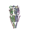
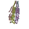
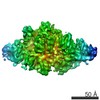
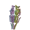
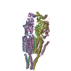
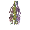
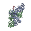
Journal: Nat Microbiol / Year: 2019 Title: An extensively glycosylated archaeal pilus survives extreme conditions. Authors: Fengbin Wang / Virginija Cvirkaite-Krupovic / Mark A B Kreutzberger / Zhangli Su / Guilherme A P de Oliveira / Tomasz Osinski / Nicholas Sherman / Frank DiMaio / Joseph S Wall / David ...Authors: Fengbin Wang / Virginija Cvirkaite-Krupovic / Mark A B Kreutzberger / Zhangli Su / Guilherme A P de Oliveira / Tomasz Osinski / Nicholas Sherman / Frank DiMaio / Joseph S Wall / David Prangishvili / Mart Krupovic / Edward H Egelman / Abstract: Pili on the surface of Sulfolobus islandicus are used for many functions, and serve as receptors for certain archaeal viruses. The cells grow optimally at pH 3 and ~80 °C, exposing these ...Pili on the surface of Sulfolobus islandicus are used for many functions, and serve as receptors for certain archaeal viruses. The cells grow optimally at pH 3 and ~80 °C, exposing these extracellular appendages to a very harsh environment. The pili, when removed from cells, resist digestion by trypsin or pepsin, and survive boiling in sodium dodecyl sulfate or 5 M guanidine hydrochloride. We used electron cryo-microscopy to determine the structure of these filaments at 4.1 Å resolution. An atomic model was built by combining the electron density map with bioinformatics without previous knowledge of the pilin sequence-an approach that should prove useful for assemblies where all of the components are not known. The atomic structure of the pilus was unusual, with almost one-third of the residues being either threonine or serine, and with many hydrophobic surface residues. While the map showed extra density consistent with glycosylation for only three residues, mass measurements suggested extensive glycosylation. We propose that this extensive glycosylation renders these filaments soluble and provides the remarkable structural stability. We also show that the overall fold of the archaeal pilin is remarkably similar to that of archaeal flagellin, establishing common evolutionary origins.
Embedding applied: NO / Shadowing applied: NO / Staining applied: NO / Vitrification applied: YES
Vitrification
Cryogen name: ETHANE / Humidity: 90 %
-
Electron microscopy imaging
Experimental equipment
Model: Titan Krios / Image courtesy: FEI Company
Microscopy
Model: FEI TITAN KRIOS
Electron gun
Electron source: FIELD EMISSION GUN / Accelerating voltage: 300 kV / Illumination mode: FLOOD BEAM
Electron lens
Mode: BRIGHT FIELD
Image recording
Average exposure time: 2 sec. / Electron dose: 48 e/Å2 / Detector mode: INTEGRATING / Film or detector model: FEI FALCON III (4k x 4k) Details: Images were stored containing 24 fractions, where each fraction corresponded to a dose of ~2 electrons per Angstrom^2.
In the structure databanks used in Yorodumi, some data are registered as the other names, "COVID-19 virus" and "2019-nCoV". Here are the details of the virus and the list of structure data.
Jan 31, 2019. EMDB accession codes are about to change! (news from PDBe EMDB page)
EMDB accession codes are about to change! (news from PDBe EMDB page)
The allocation of 4 digits for EMDB accession codes will soon come to an end. Whilst these codes will remain in use, new EMDB accession codes will include an additional digit and will expand incrementally as the available range of codes is exhausted. The current 4-digit format prefixed with “EMD-” (i.e. EMD-XXXX) will advance to a 5-digit format (i.e. EMD-XXXXX), and so on. It is currently estimated that the 4-digit codes will be depleted around Spring 2019, at which point the 5-digit format will come into force.
The EM Navigator/Yorodumi systems omit the EMD- prefix.
Related info.:Q: What is EMD? / ID/Accession-code notation in Yorodumi/EM Navigator
Yorodumi is a browser for structure data from EMDB, PDB, SASBDB, etc.
This page is also the successor to EM Navigator detail page, and also detail information page/front-end page for Omokage search.
The word "yorodu" (or yorozu) is an old Japanese word meaning "ten thousand". "mi" (miru) is to see.
Related info.:EMDB / PDB / SASBDB / Comparison of 3 databanks / Yorodumi Search / Aug 31, 2016. New EM Navigator & Yorodumi / Yorodumi Papers / Jmol/JSmol / Function and homology information / Changes in new EM Navigator and Yorodumi
 Movie
Movie Controller


 Open data
 Basic information
 Components
 Keywords
 Function and homology information
 Sulfolobus islandicus LAL14/1 (archaea)
 Authors
 United States, 1items
  Citation
 Structure visualization
 Downloads & links
 Other downloads
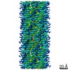
 PDBj
PDBj Assembly
 Sample preparation
 Electron microscopy imaging
 FIELD EMISSION GUN / Accelerating voltage: 300 kV / Illumination mode: FLOOD BEAM
 Processing