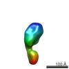
Movie
Movie Controller

[English] 日本語
 Yorodumi
- EMDB-21455: Cryo-EM structure of filamentous PFD from Methanocaldococcus jann... -
+ Open data

- Basic information
| Entry | Database: EMDB / ID: EMD-21455 | ||||||||||||
|---|---|---|---|---|---|---|---|---|---|---|---|---|---|
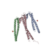
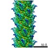
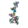
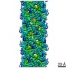
| Title | Cryo-EM structure of filamentous PFD from Methanocaldococcus jannaschii | ||||||||||||
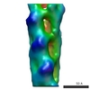
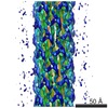
 Map data Map data | Cryo-EM structure of filamentous PFD | ||||||||||||
 Sample Sample |
| ||||||||||||
 Keywords Keywords | helical symmetry / nanowire / prefoldin / PROTEIN FIBRIL | ||||||||||||
| Function / homology | Prefoldin alpha-like / Prefoldin alpha subunit, archaea-type / Prefoldin subunit / prefoldin complex / Prefoldin / : / protein folding / cytoplasm / Prefoldin subunit alpha 2 Function and homology information Function and homology information | ||||||||||||
| Biological species |   Methanocaldococcus jannaschii (archaea) / Methanocaldococcus jannaschii (strain ATCC 43067 / DSM 2661 / JAL-1 / JCM 10045 / NBRC 100440) (archaea) Methanocaldococcus jannaschii (archaea) / Methanocaldococcus jannaschii (strain ATCC 43067 / DSM 2661 / JAL-1 / JCM 10045 / NBRC 100440) (archaea) | ||||||||||||
| Method | helical reconstruction / cryo EM / Resolution: 6.0 Å | ||||||||||||
 Authors Authors | Wang F / Chen YX | ||||||||||||
| Funding support |  United States, 3 items United States, 3 items
| ||||||||||||
 Citation Citation | Journal: ACS Nano / Year: 2020 Title: Structural Determination of a Filamentous Chaperone to Fabricate Electronically Conductive Metalloprotein Nanowires. Authors: Yun X Chen / Nicole L Ing / Fengbin Wang / Dawei Xu / Nancy B Sloan / Nga T Lam / Daniel L Winter / Edward H Egelman / Allon I Hochbaum / Douglas S Clark / Dominic J Glover /  Abstract: The transfer of electrons through protein complexes is central to cellular respiration. Exploiting proteins for charge transfer in a controllable fashion has the potential to revolutionize the ...The transfer of electrons through protein complexes is central to cellular respiration. Exploiting proteins for charge transfer in a controllable fashion has the potential to revolutionize the integration of biological systems and electronic devices. Here we characterize the structure of an ultrastable protein filament and engineer the filament subunits to create electronically conductive nanowires under aqueous conditions. Cryoelectron microscopy was used to resolve the helical structure of gamma-prefoldin, a filamentous protein from a hyperthermophilic archaeon. Conjugation of tetra-heme c3-type cytochromes along the longitudinal axis of the filament created nanowires capable of long-range electron transfer. Electrochemical transport measurements indicated networks of the nanowires capable of conducting current between electrodes at the redox potential of the cytochromes. Functionalization of these highly engineerable nanowires with other molecules, such as redox enzymes, may be useful for bioelectronic applications. | ||||||||||||
| History |
|
- Structure visualization
| Movie |
Movie viewer |
|---|---|
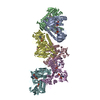
| Structure viewer | EM map: SurfViewMolmilJmol/JSmol |
| Supplemental images |

- Downloads & links
-EMDB archive
| Map data | emd_21455.map.gz | 3.4 MB | EMDB map data format | |
|---|---|---|---|---|
| Header (meta data) | emd-21455-v30.xmlemd-21455.xml | 14.5 KB 14.5 KB | Display Display | EMDB header |
| Images | 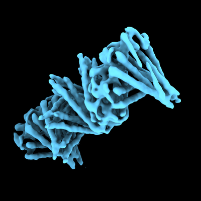 emd_21455.png emd_21455.png | 86.8 KB | ||
| Filedesc metadata | emd-21455.cif.gz | 5.8 KB | ||
| Archive directory |  http://ftp.pdbj.org/pub/emdb/structures/EMD-21455ftp://ftp.pdbj.org/pub/emdb/structures/EMD-21455 http://ftp.pdbj.org/pub/emdb/structures/EMD-21455ftp://ftp.pdbj.org/pub/emdb/structures/EMD-21455 | HTTPS FTP |
-Related structure data
| Related structure data | 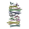 6vy1MC M: atomic model generated by this map C: citing same article ( |
|---|---|
| Similar structure data |

-Links
| EMDB pages | EMDB (EBI/PDBe) / EMDataResource |
|---|---|
| Related items in Molecule of the Month |

-Map
| File | Download / File: emd_21455.map.gz / Format: CCP4 / Size: 216 MB / Type: IMAGE STORED AS FLOATING POINT NUMBER (4 BYTES) | ||||||||||||||||||||||||||||||||||||||||||||||||||||||||||||
|---|---|---|---|---|---|---|---|---|---|---|---|---|---|---|---|---|---|---|---|---|---|---|---|---|---|---|---|---|---|---|---|---|---|---|---|---|---|---|---|---|---|---|---|---|---|---|---|---|---|---|---|---|---|---|---|---|---|---|---|---|---|
| Annotation | Cryo-EM structure of filamentous PFD | ||||||||||||||||||||||||||||||||||||||||||||||||||||||||||||
| Projections & slices | Image control
Images are generated by Spider. | ||||||||||||||||||||||||||||||||||||||||||||||||||||||||||||
| Voxel size | X=Y=Z: 1.09 Å | ||||||||||||||||||||||||||||||||||||||||||||||||||||||||||||
| Density |
| ||||||||||||||||||||||||||||||||||||||||||||||||||||||||||||
| Symmetry | Space group: 1 | ||||||||||||||||||||||||||||||||||||||||||||||||||||||||||||
| Details | EMDB XML:
CCP4 map header:
| ||||||||||||||||||||||||||||||||||||||||||||||||||||||||||||
 Z (Sec.)
 Y (Row.)
 X (Col.)


-Supplemental data
- Sample components
-Entire : Methanocaldococcus jannaschii gamma-prefoldin
| Entire | Name: Methanocaldococcus jannaschii gamma-prefoldin |
|---|---|
| Components |
|
-Supramolecule #1: Methanocaldococcus jannaschii gamma-prefoldin
| Supramolecule | Name: Methanocaldococcus jannaschii gamma-prefoldin / type: complex / ID: 1 / Parent: 0 / Macromolecule list: all |
|---|---|
| Source (natural) | Organism: Methanocaldococcus jannaschii (archaea) |
-Macromolecule #1: Prefoldin subunit alpha 2
| Macromolecule | Name: Prefoldin subunit alpha 2 / type: protein_or_peptide / ID: 1 / Number of copies: 14 / Enantiomer: LEVO |
|---|---|
| Source (natural) | Organism: Methanocaldococcus jannaschii (strain ATCC 43067 / DSM 2661 / JAL-1 / JCM 10045 / NBRC 100440) (archaea) Strain: ATCC 43067 / DSM 2661 / JAL-1 / JCM 10045 / NBRC 100440 |
| Molecular weight | Theoretical: 16.410641 KDa |
| Recombinant expression | Organism:  |
| Sequence | String: MVNEVIDINE AVRAYIAQIE GLRAEIGRLD ATIATLRQSL ATLKSLKTLG EGKTVLVPVG SIAQVEMKVE KMDKVVVSVG QNISAELEY EEALKYIEDE IKKLLTFRLV LEQAIAELYA KIEDLIAEAQ QTSEEEKAEE EENEEKAE UniProtKB: Prefoldin subunit alpha 2 |
-Experimental details
-Structure determination
| Method | cryo EM |
|---|---|
 Processing Processing | helical reconstruction |
| Aggregation state | filament |
-Sample preparation
| Buffer | pH: 7.5 |
|---|---|
| Vitrification | Cryogen name: ETHANE |
- Electron microscopy
| Microscope | FEI TITAN KRIOS |
|---|---|
| Image recording | Film or detector model: FEI FALCON III (4k x 4k) / Average electron dose: 55.0 e/Å2 |
| Electron beam | Acceleration voltage: 300 kV / Electron source:  FIELD EMISSION GUN FIELD EMISSION GUN |
| Electron optics | Illumination mode: FLOOD BEAM / Imaging mode: BRIGHT FIELD |
| Experimental equipment |  Model: Titan Krios / Image courtesy: FEI Company |
-Image processing
| Final reconstruction | Applied symmetry - Helical parameters - Δz: 18.27 Å Applied symmetry - Helical parameters - Δ&Phi: -48.93 ° Applied symmetry - Helical parameters - Axial symmetry: C1 (asymmetric) Resolution.type: BY AUTHOR / Resolution: 6.0 Å / Resolution method: OTHER / Details: d99, model:map FSC and map:map FSC / Number images used: 32227 |
|---|---|
| CTF correction | Type: PHASE FLIPPING AND AMPLITUDE CORRECTION |
| Startup model | Type of model: OTHER / Details: featureless cylinder |
| Final angle assignment | Type: NOT APPLICABLE |