 Movie
Movie Controller

[English] 日本語
 Yorodumi
- PDB-3eao: Crystal structure of recombinant rat selenoprotein thioredoxin re... -
+ Open data

- Basic information
| Entry | Database: PDB / ID: 3eao | ||||||
|---|---|---|---|---|---|---|---|
| Title | Crystal structure of recombinant rat selenoprotein thioredoxin reductase 1 with oxidized C-terminal tail | ||||||
 Components Components | Thioredoxin reductase 1, cytoplasmic | ||||||
 Keywords Keywords | OXIDOREDUCTASE / Selenoprotein / mammalian thioredoxin reductase 1 / selenocysteine / selenenylsulfide / FAD / Flavoprotein / NADP / Phosphoprotein / Redox-active center / Selenium | ||||||
| Function / homology |  Function and homology information Function and homology informationselenate reductase activity / halogen metabolic process / Metabolism of ingested MeSeO2H into MeSeH / benzene-containing compound metabolic process / Interconversion of nucleotide di- and triphosphates / : / Detoxification of Reactive Oxygen Species / NADPH peroxidase / NADPH peroxidase activity / cellular response to hyperoxia ...selenate reductase activity / halogen metabolic process / Metabolism of ingested MeSeO2H into MeSeH / benzene-containing compound metabolic process / Interconversion of nucleotide di- and triphosphates / : / Detoxification of Reactive Oxygen Species / NADPH peroxidase / NADPH peroxidase activity / cellular response to hyperoxia / TP53 Regulates Metabolic Genes / mercury ion binding / NAD(P)H oxidase H2O2-forming activity / thioredoxin-disulfide reductase (NADPH) / response to selenium ion / thioredoxin-disulfide reductase (NADPH) activity / mesoderm formation / response to hyperoxia / response to axon injury / gastrulation / FAD binding / cell redox homeostasis / cellular response to copper ion / hydrogen peroxide catabolic process / fibrillar center / response to oxidative stress / cell population proliferation / response to xenobiotic stimulus / positive regulation of apoptotic process / neuronal cell body / mitochondrion / nucleoplasm / identical protein binding / nucleus / cytosol / cytoplasm Similarity search - Function | ||||||
| Biological species |  | ||||||
| Method |  X-RAY DIFFRACTION / SYNCHROTRON / MOLECULAR REPLACEMENT / Resolution: 3.1 Å X-RAY DIFFRACTION / SYNCHROTRON / MOLECULAR REPLACEMENT / Resolution: 3.1 Å | ||||||
 Authors Authors | Sandalova, T. / Cheng, Q. / Lindqvist, Y. / Arner, E. | ||||||
 Citation Citation | Journal: J.Biol.Chem. / Year: 2009 Title: Crystal structure and catalysis of the selenoprotein thioredoxin reductase 1. Authors: Cheng, Q. / Sandalova, T. / Lindqvist, Y. / Arner, E.S. #1: Journal: J.Mol.Biol. / Year: 1999 Title: High-level expression in Escherichia coli of selenocysteine-containing rat thioredoxin reductase utilizing gene fusions with engineered bacterial-type SECIS elements and co-expression with the ...Title: High-level expression in Escherichia coli of selenocysteine-containing rat thioredoxin reductase utilizing gene fusions with engineered bacterial-type SECIS elements and co-expression with the selA, selB and selC genes. Authors: Arner, E.S. / Sarioglu, H. / Lottspeich, F. / Holmgren, A. / Bock, A. | ||||||
| History |
|
- Structure visualization
| Structure viewer | Molecule: MolmilJmol/JSmol |
|---|
- Downloads & links
-Download
| PDBx/mmCIF format | 3eao.cif.gz | 562.9 KB | Display | PDBx/mmCIF format |
|---|---|---|---|---|
| PDB format | pdb3eao.ent.gz | 463.6 KB | Display | PDB format |
| PDBx/mmJSON format | 3eao.json.gz | Tree view | PDBx/mmJSON format | |
| Others |  Other downloads Other downloads |
-Validation report
| Arichive directory | https://data.pdbj.org/pub/pdb/validation_reports/ea/3eaoftp://data.pdbj.org/pub/pdb/validation_reports/ea/3eao | HTTPS FTP |
|---|
-Related structure data
| Related structure data |  3eanSC S: Starting model for refinement C: citing same article ( |
|---|---|
| Similar structure data |


-Links
 PDBj
PDBj


- Assembly
| Deposited unit | 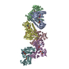
| ||||||||||||||||||||||||||||||||||||||||||||||||||||||||||||||||||||||||||||||||||||||||||||||||||||||||||||||||||||||||||||||||||||||||||||||||||||||||||||||||||||||||||||||||||||||||||||||||
|---|---|---|---|---|---|---|---|---|---|---|---|---|---|---|---|---|---|---|---|---|---|---|---|---|---|---|---|---|---|---|---|---|---|---|---|---|---|---|---|---|---|---|---|---|---|---|---|---|---|---|---|---|---|---|---|---|---|---|---|---|---|---|---|---|---|---|---|---|---|---|---|---|---|---|---|---|---|---|---|---|---|---|---|---|---|---|---|---|---|---|---|---|---|---|---|---|---|---|---|---|---|---|---|---|---|---|---|---|---|---|---|---|---|---|---|---|---|---|---|---|---|---|---|---|---|---|---|---|---|---|---|---|---|---|---|---|---|---|---|---|---|---|---|---|---|---|---|---|---|---|---|---|---|---|---|---|---|---|---|---|---|---|---|---|---|---|---|---|---|---|---|---|---|---|---|---|---|---|---|---|---|---|---|---|---|---|---|---|---|---|---|---|---|
| 1 | 
| ||||||||||||||||||||||||||||||||||||||||||||||||||||||||||||||||||||||||||||||||||||||||||||||||||||||||||||||||||||||||||||||||||||||||||||||||||||||||||||||||||||||||||||||||||||||||||||||||
| 2 | 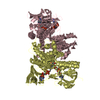
| ||||||||||||||||||||||||||||||||||||||||||||||||||||||||||||||||||||||||||||||||||||||||||||||||||||||||||||||||||||||||||||||||||||||||||||||||||||||||||||||||||||||||||||||||||||||||||||||||
| 3 | 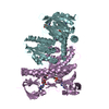
| ||||||||||||||||||||||||||||||||||||||||||||||||||||||||||||||||||||||||||||||||||||||||||||||||||||||||||||||||||||||||||||||||||||||||||||||||||||||||||||||||||||||||||||||||||||||||||||||||
| Unit cell |
| ||||||||||||||||||||||||||||||||||||||||||||||||||||||||||||||||||||||||||||||||||||||||||||||||||||||||||||||||||||||||||||||||||||||||||||||||||||||||||||||||||||||||||||||||||||||||||||||||
| Noncrystallographic symmetry (NCS) | NCS domain:
NCS domain segments:
NCS ensembles :
| ||||||||||||||||||||||||||||||||||||||||||||||||||||||||||||||||||||||||||||||||||||||||||||||||||||||||||||||||||||||||||||||||||||||||||||||||||||||||||||||||||||||||||||||||||||||||||||||||
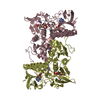
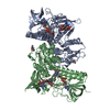
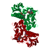
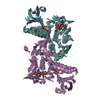
| Details | The biological assembly is a dimer, the unit cell contains three dimers |
-Components
| #1: Protein | Mass: 54736.293 Da / Num. of mol.: 6 Source method: isolated from a genetically manipulated source Details: residue 426 is Ile / Source: (gene. exp.)  References: UniProt: O89049, thioredoxin-disulfide reductase (NADPH) #2: Chemical | ChemComp-FAD /   Mass: 785.550 Da / Num. of mol.: 6 / Source method: obtained synthetically / Formula: C27H33N9O15P2 / Comment: FAD*YM Mass: 785.550 Da / Num. of mol.: 6 / Source method: obtained synthetically / Formula: C27H33N9O15P2 / Comment: FAD*YM#3: Chemical | ChemComp-NAP /   Mass: 743.405 Da / Num. of mol.: 6 / Source method: obtained synthetically / Formula: C21H28N7O17P3 Mass: 743.405 Da / Num. of mol.: 6 / Source method: obtained synthetically / Formula: C21H28N7O17P3#4: Water | ChemComp-HOH / |  Mass: 18.015 Da / Num. of mol.: 36 / Source method: isolated from a natural source / Formula: H2O Mass: 18.015 Da / Num. of mol.: 36 / Source method: isolated from a natural source / Formula: H2OHas protein modification | Y | Sequence details | THE SEQUENCE MATCHES WITH NP_113802 FROM NCBI DATABASE | |
|---|
-Experimental details
-Experiment
| Experiment | Method: X-RAY DIFFRACTION / Number of used crystals: 1 |
|---|
- Sample preparation
| Crystal | Density Matthews: 2.77 Å3/Da / Density % sol: 55.52 % |
|---|---|
| Crystal grow | Temperature: 298 K / Method: vapor diffusion, hanging drop / pH: 7.5 Details: 0.1M HEPES, PEG 3350 15%, 12% of ethylene glycol, pH 7.5, VAPOR DIFFUSION, HANGING DROP, temperature 298K |
-Data collection
| Diffraction | Mean temperature: 100 K |
|---|---|
| Diffraction source | Source: SYNCHROTRON / Site: MAX II  / Beamline: I911-3 / Wavelength: 0.908 Å / Beamline: I911-3 / Wavelength: 0.908 Å |
| Detector | Type: MARMOSAIC 225 mm CCD / Detector: CCD / Date: Nov 6, 2007 / Details: mirrors |
| Radiation | Monochromator: Double crystal monochromator, Si(111) / Protocol: SINGLE WAVELENGTH / Monochromatic (M) / Laue (L): M / Scattering type: x-ray |
| Radiation wavelength | Wavelength: 0.908 Å / Relative weight: 1 |
| Reflection | Resolution: 3.1→64 Å / Num. all: 64804 / Num. obs: 64804 / % possible obs: 99.8 % / Observed criterion σ(F): 0 / Observed criterion σ(I): 0 / Redundancy: 3.3 % / Biso Wilson estimate: 76 Å2 / Rmerge(I) obs: 0.131 / Rsym value: 0.131 / Net I/σ(I): 10.9 |
| Reflection shell | Resolution: 3.101→3.27 Å / Redundancy: 3 % / Rmerge(I) obs: 0.559 / Mean I/σ(I) obs: 1.9 / % possible all: 100 |
- Processing
| Software |
| |||||||||||||||||||||||||||||||||||||||||||||||||||||||||||||||||||||||||||||||||||||||||||||||||||||||||||||||||||||||||||||||||||||||||||||||||||||||||||||||||||||||||||||||
|---|---|---|---|---|---|---|---|---|---|---|---|---|---|---|---|---|---|---|---|---|---|---|---|---|---|---|---|---|---|---|---|---|---|---|---|---|---|---|---|---|---|---|---|---|---|---|---|---|---|---|---|---|---|---|---|---|---|---|---|---|---|---|---|---|---|---|---|---|---|---|---|---|---|---|---|---|---|---|---|---|---|---|---|---|---|---|---|---|---|---|---|---|---|---|---|---|---|---|---|---|---|---|---|---|---|---|---|---|---|---|---|---|---|---|---|---|---|---|---|---|---|---|---|---|---|---|---|---|---|---|---|---|---|---|---|---|---|---|---|---|---|---|---|---|---|---|---|---|---|---|---|---|---|---|---|---|---|---|---|---|---|---|---|---|---|---|---|---|---|---|---|---|---|---|---|---|
| Refinement | Method to determine structure: MOLECULAR REPLACEMENT Starting model: PDB ENTRY 3EAN Resolution: 3.1→30 Å / Cor.coef. Fo:Fc: 0.87 / Cor.coef. Fo:Fc free: 0.83 / SU B: 26.935 / SU ML: 0.46 / Isotropic thermal model: isotropic / Cross valid method: THROUGHOUT / σ(F): 0 / σ(I): 0 / ESU R Free: 0.561 / Stereochemistry target values: MAXIMUM LIKELIHOOD / Details: HYDROGENS HAVE BEEN ADDED IN THE RIDING POSITIONS
| |||||||||||||||||||||||||||||||||||||||||||||||||||||||||||||||||||||||||||||||||||||||||||||||||||||||||||||||||||||||||||||||||||||||||||||||||||||||||||||||||||||||||||||||
| Solvent computation | Ion probe radii: 0.8 Å / Shrinkage radii: 0.8 Å / VDW probe radii: 1.2 Å / Solvent model: MASK | |||||||||||||||||||||||||||||||||||||||||||||||||||||||||||||||||||||||||||||||||||||||||||||||||||||||||||||||||||||||||||||||||||||||||||||||||||||||||||||||||||||||||||||||
| Displacement parameters | Biso mean: 56.314 Å2
| |||||||||||||||||||||||||||||||||||||||||||||||||||||||||||||||||||||||||||||||||||||||||||||||||||||||||||||||||||||||||||||||||||||||||||||||||||||||||||||||||||||||||||||||
| Refinement step | Cycle: LAST / Resolution: 3.1→30 Å
| |||||||||||||||||||||||||||||||||||||||||||||||||||||||||||||||||||||||||||||||||||||||||||||||||||||||||||||||||||||||||||||||||||||||||||||||||||||||||||||||||||||||||||||||
| Refine LS restraints |
| |||||||||||||||||||||||||||||||||||||||||||||||||||||||||||||||||||||||||||||||||||||||||||||||||||||||||||||||||||||||||||||||||||||||||||||||||||||||||||||||||||||||||||||||
| Refine LS restraints NCS | Refine-ID: X-RAY DIFFRACTION
| |||||||||||||||||||||||||||||||||||||||||||||||||||||||||||||||||||||||||||||||||||||||||||||||||||||||||||||||||||||||||||||||||||||||||||||||||||||||||||||||||||||||||||||||
| LS refinement shell | Resolution: 3.1→3.18 Å / Total num. of bins used: 20
|
