 Movie
Movie Controller


+ Open data

- Basic information
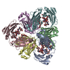
| Entry | Database: PDB / ID: 2zdi | ||||||
|---|---|---|---|---|---|---|---|
| Title | Crystal structure of Prefoldin from Pyrococcus horikoshii OT3 | ||||||
 Components Components |
| ||||||
 Keywords Keywords | CHAPERONE / PREFOLDIN / Cytoplasm | ||||||
| Function / homology |  Function and homology information Function and homology informationprefoldin complex / protein folding chaperone / : / protein folding / cytoplasm Similarity search - Function | ||||||
| Biological species |   Pyrococcus horikoshii (archaea) Pyrococcus horikoshii (archaea) | ||||||
| Method |  X-RAY DIFFRACTION / SYNCHROTRON / MOLECULAR REPLACEMENT / Resolution: 3 Å X-RAY DIFFRACTION / SYNCHROTRON / MOLECULAR REPLACEMENT / Resolution: 3 Å | ||||||
 Authors Authors | Kida, H. / Miki, K. | ||||||
 Citation Citation | Journal: J.Mol.Biol. / Year: 2008 Title: Structure and molecular dynamics simulation of archaeal prefoldin: the molecular mechanism for binding and recognition of nonnative substrate proteins Authors: Ohtaki, A. / Kida, H. / Miyata, Y. / Ide, N. / Yonezawa, A. / Arakawa, T. / Iizuka, R. / Noguchi, K. / Kita, A. / Odaka, M. / Miki, K. / Yohda, M. | ||||||
| History |
|
- Structure visualization
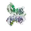
| Structure viewer | Molecule: MolmilJmol/JSmol |
|---|
- Downloads & links
-Download
| PDBx/mmCIF format | 2zdi.cif.gz | 78.6 KB | Display | PDBx/mmCIF format |
|---|---|---|---|---|
| PDB format | pdb2zdi.ent.gz | 59.1 KB | Display | PDB format |
| PDBx/mmJSON format | 2zdi.json.gz | Tree view | PDBx/mmJSON format | |
| Others |  Other downloads Other downloads |
-Validation report
| Arichive directory | https://data.pdbj.org/pub/pdb/validation_reports/zd/2zdiftp://data.pdbj.org/pub/pdb/validation_reports/zd/2zdi | HTTPS FTP |
|---|
-Related structure data
| Related structure data | 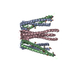 1fxkS S: Starting model for refinement |
|---|---|
| Similar structure data |

-Links
 PDBj
PDBj
- Assembly
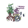
| Deposited unit | 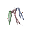
| ||||||||
|---|---|---|---|---|---|---|---|---|---|
| 1 | 
| ||||||||
| Unit cell |
|
-Components
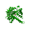
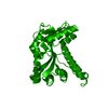
| #1: Protein | Mass: 13333.361 Da / Num. of mol.: 2 Source method: isolated from a genetically manipulated source Source: (gene. exp.) Pyrococcus horikoshii (archaea) / Gene: pfdB / Plasmid: pET23b / Production host:  #2: Protein | | Mass: 17027.660 Da / Num. of mol.: 1 Source method: isolated from a genetically manipulated source Source: (gene. exp.) Pyrococcus horikoshii (archaea) / Gene: pfdA / Plasmid: pET23b / Production host: #3: Chemical |   Mass: 96.063 Da / Num. of mol.: 3 / Source method: obtained synthetically / Formula: SO4 Mass: 96.063 Da / Num. of mol.: 3 / Source method: obtained synthetically / Formula: SO4Sequence details | THE N-TERMINAL 3 RESIDUES (MET, ILE, ARG) OF CHAIN C (ENTITY 2) WERE INCLUDED IN ACCESSION NO. ...THE N-TERMINAL 3 RESIDUES (MET, ILE, ARG) OF CHAIN C (ENTITY 2) WERE INCLUDED IN ACCESSION NO. BA000001-547 OF DDBJ DATABASE, BUT NOT IN ACCESSION NO. O58263 OF UNIPROT DATABASE. | |
|---|
-Experimental details
-Experiment
| Experiment | Method: X-RAY DIFFRACTION / Number of used crystals: 1 |
|---|
- Sample preparation
| Crystal | Density Matthews: 2.89 Å3/Da / Density % sol: 57.51 % |
|---|---|
| Crystal grow | Temperature: 291 K / Method: vapor diffusion, sitting drop / pH: 6.5 Details: 30% PEG 400, 0.1M Sodium chloride, 0.1M Lithium sulfate, 0.1M MES-NaOH pH6.5, VAPOR DIFFUSION, SITTING DROP, temperature 291K |
-Data collection
| Diffraction | Mean temperature: 100 K |
|---|---|
| Diffraction source | Source: SYNCHROTRON / Site: Photon Factory  / Beamline: AR-NW12A / Wavelength: 1 Å / Beamline: AR-NW12A / Wavelength: 1 Å |
| Detector | Type: ADSC QUANTUM 210 / Detector: CCD / Date: Oct 25, 2005 |
| Radiation | Protocol: SINGLE WAVELENGTH / Monochromatic (M) / Laue (L): M / Scattering type: x-ray |
| Radiation wavelength | Wavelength: 1 Å / Relative weight: 1 |
| Reflection | Resolution: 3→50 Å / Num. obs: 10215 / % possible obs: 94.1 % / Observed criterion σ(I): -3 / Biso Wilson estimate: 66.56 Å2 / Rsym value: 0.055 / Net I/σ(I): 25.2 |
- Processing
| Software |
| ||||||||||||||||||||
|---|---|---|---|---|---|---|---|---|---|---|---|---|---|---|---|---|---|---|---|---|---|
| Refinement | Method to determine structure: MOLECULAR REPLACEMENT Starting model: PDB ENTRY 1FXK Resolution: 3→50 Å / Cross valid method: THROUGHOUT / σ(I): -3
| ||||||||||||||||||||
| Displacement parameters | Biso mean: 59.14 Å2
| ||||||||||||||||||||
| Refinement step | Cycle: LAST / Resolution: 3→50 Å
|
