 ムービー
ムービー コントローラー
 構造ビューア
 EMN検索について


-検索条件
-検索結果
検索 (著者・登録者: s. & wang)の結果1,073件中、1から50件目までを表示しています
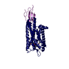
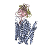
PDB-8e6q: 
Human TRPM2 ion channel in 1 mM ADPR
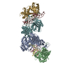
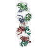
PDB-8e6r:
Human TRPM2 ion channel in 1 mM dADPR
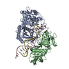
PDB-8e6s:
Human TRPM2 ion channel in 1 mM dADPR and Ca2+
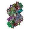
PDB-8e6t:
Human TRPM2 ion channel in 1 mM BR-ADPR
PDB-8e6u:
Human TRPM2 ion channel in 1 mM F-dADPR
PDB-8e6v:
MHR1/2 and NUDT9H of human TRPM2 in 1 mM dADPR (local refinement)

PDB-8tox:
Cryo-EM structure of BG505 Env mutant A517E in complex with antibody ACS202 Fab
PDB-8xva:
Human TOM complex with whole Tom20
PDB-8vww:
CCHFV GP38 bound to ADI-46152 and ADI-58048 Fabs
PDB-8k58:
The cryo-EM map of close TIEA-TIC complex

PDB-8k5a:
The cryo-EM map of open TIEA-TIC complex
PDB-8wlu:
Cryo-EM structure of bat RsSHC014 spike glycoprotein
PDB-8wly:
Cryo-EM structure of bat WIV1 spike glycoprotein
PDB-8wlz:
Cryo-EM structure of the WIV1 S-hACE2 complex

PDB-8wq0:
Cryo-EM structure of WIV1 spike glycoprotein (the closed state)
PDB-9bjk:
Inactive mu opioid receptor bound to Nb6, naloxone and NAM
PDB-8k87:
Dimer structure of procaryotic Ago
PDB-8k88:
Structure of procaryotic Ago
PDB-8tow:
Structure of a mutated photosystem II complex reveals perturbation of the oxygen-evolving complex
PDB-8khr:
Cryo-EM structure of EBV gH/gL-gp42 in complex with fab 2C1

PDB-8p2w:
Structure of human SIT1 (focussed map / refinement)

PDB-8p2x:
Structure of human SIT1:ACE2 complex (open PD conformation)

PDB-8p2y:
Structure of human SIT1:ACE2 complex (closed PD conformation)

PDB-8p2z:
Structure of human SIT1 bound to L-pipecolate (focussed map / refinement)
PDB-8p30:
Structure of human SIT1:ACE2 complex (open PD conformation) bound to L-pipecolate

PDB-8p31:
Structure of human SIT1:ACE2 complex (closed PD conformation) bound to L-pipecolate

PDB-8ouo:
Human TPC2 in Complex with Antagonist (S)-SG-094

PDB-8hat:
NARROW LEAF 1-open from Japonica

PDB-8ikg:
Cryo-EM structure of human receptor with G proteins
PDB-8ikh:
Cryo-EM structure of human receptor with G proteins

PDB-8iyq:
Structure of CbCas9 bound to 20-nucleotide complementary DNA substrate

PDB-8wmh:
Structure of CbCas9 bound to 6-nucleotide complementary DNA substrate

PDB-8wmm:
Structure of CbCas9-PcrIIC1 complex bound to 28-bp DNA substrate (20-nt complementary)

PDB-8wmn:
Structure of CbCas9-PcrIIC1 complex bound to 62-bp DNA substrate (symmetric 20-nt complementary)

PDB-8wr4:
Structure of CbCas9-PcrIIC1 complex bound to 62-bp DNA substrate (non-targeting complex)

PDB-8xbs:
C. elegans apo-SID1 structure

PDB-8xc1:
C. elegans SID1 in complex with dsRNA

PDB-8jj1:
Cryo-EM structure of GluN1-2A NMDAR in complex with human Fab2G7 in two fab conformation
PDB-8hau:
NARROW LEAF 1 from Indica

PDB-8jj2:
Cryo-EM structure of GluN1-2A NMDAR in complex with human Fab2G7 in one fab conformation

PDB-8p2e:
Homotypic interacting B1 fab bound to Chondroitin Sulfate A

PDB-8jj0:
Cryo-EM structure of GluN1-2A NMDAR in complex with human Fab5F6 in one fab bind conformation

PDB-8jiz:
Cryo-EM structure of GluN1-2A NMDAR in complex with human Fab5F6 in two fab bind conformation

PDB-8tte:
Protonated state of NorA at pH 5.0
PDB-8ttf:
NorA double mutant - E222QD307N at pH 7.5

PDB-8ttg:
NorA single mutant - E222Q at pH 7.5

PDB-8tth:
NorA single mutant - D307N at pH 7.5

PDB-8x8q:
Structure of enterovirus protease in complex host factor

PDB-8vue:
L5A7 Fab bound to Indonesia2005 Hemagglutinin
PDB-8vuz:
L5A7 Fab bound to 28H6E11 anti-idiotype Fab
ページ:
 wwPDBはEMDBデータモデルのバージョン3へ移行します
