ムービー
ムービー コントローラー


+ データを開く

- 基本情報
基本情報
| 登録情報 | データベース: PDB / ID: 8x8q | |||||||||||||||||||||
|---|---|---|---|---|---|---|---|---|---|---|---|---|---|---|---|---|---|---|---|---|---|---|
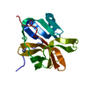
| タイトル | Structure of enterovirus protease in complex host factor | |||||||||||||||||||||
 要素 要素 |
| |||||||||||||||||||||
 キーワード キーワード | CELL CYCLE / host protein / VIRAL PROTEIN / CELL INVASION | |||||||||||||||||||||
| 機能・相同性 |  機能・相同性情報 機能・相同性情報peptidyl-histidine methylation / regulation of uterine smooth muscle contraction / protein-histidine N-methyltransferase / protein-L-histidine N-tele-methyltransferase activity / actin modification / histone H3K36 methyltransferase activity / histone H3K4 methyltransferase activity / positive regulation of muscle cell differentiation / cysteine-type peptidase activity / symbiont-mediated suppression of host mRNA export from nucleus ...peptidyl-histidine methylation / regulation of uterine smooth muscle contraction / protein-histidine N-methyltransferase / protein-L-histidine N-tele-methyltransferase activity / actin modification / histone H3K36 methyltransferase activity / histone H3K4 methyltransferase activity / positive regulation of muscle cell differentiation / cysteine-type peptidase activity / symbiont-mediated suppression of host mRNA export from nucleus / symbiont genome entry into host cell via pore formation in plasma membrane / T=pseudo3 icosahedral viral capsid / helicase activity / host cell cytoplasmic vesicle membrane / PKMTs methylate histone lysines / channel activity / actin binding / monoatomic ion transmembrane transport / RNA polymerase II-specific DNA-binding transcription factor binding / transcription coactivator activity / symbiont-mediated suppression of host innate immune response / endocytosis involved in viral entry into host cell / symbiont-mediated activation of host autophagy / RNA-directed RNA polymerase activity / positive regulation of DNA-templated transcription / virion attachment to host cell / chromatin / host cell nucleus / positive regulation of transcription by RNA polymerase II / proteolysis / RNA binding / zinc ion binding / nucleoplasm / ATP binding / membrane / cytoplasm 類似検索 - 分子機能 | |||||||||||||||||||||
| 生物種 |  Homo sapiens (ヒト) Homo sapiens (ヒト)  Enterovirus A71 (エンテロウイルス) Enterovirus A71 (エンテロウイルス) | |||||||||||||||||||||
| 手法 | 電子顕微鏡法 / 単粒子再構成法 / クライオ電子顕微鏡法 / 解像度: 3.14 Å | |||||||||||||||||||||
 データ登録者 データ登録者 | Gao, X. / Cui, S. | |||||||||||||||||||||
| 資金援助 | 1件
| |||||||||||||||||||||
 引用 引用 | ジャーナル: Nat Commun / 年: 2024 タイトル: The EV71 2A protease occupies the central cleft of SETD3 and disrupts SETD3-actin interaction. 著者: Xiaopan Gao / Bei Wang / Kaixiang Zhu / Linyue Wang / Bo Qin / Kun Shang / Wei Ding / Jianwei Wang / Sheng Cui /  要旨: SETD3 is an essential host factor for the replication of a variety of enteroviruses that specifically interacts with viral protease 2A. However, the interaction between SETD3 and the 2A protease has ...SETD3 is an essential host factor for the replication of a variety of enteroviruses that specifically interacts with viral protease 2A. However, the interaction between SETD3 and the 2A protease has not been fully characterized. Here, we use X-ray crystallography and cryo-electron microscopy to determine the structures of SETD3 complexed with the 2A protease of EV71 to 3.5 Å and 3.1 Å resolution, respectively. We find that the 2A protease occupies the V-shaped central cleft of SETD3 through two discrete sites. The relative positions of the two proteins vary in the crystal and cryo-EM structures, showing dynamic binding. A biolayer interferometry assay shows that the EV71 2A protease outcompetes actin for SETD3 binding. We identify key 2A residues involved in SETD3 binding and demonstrate that 2A's ability to bind SETD3 correlates with EV71 production in cells. Coimmunoprecipitation experiments in EV71 infected and 2A expressing cells indicate that 2A interferes with the SETD3-actin complex, and the disruption of this complex reduces enterovirus replication. Together, these results reveal the molecular mechanism underlying the interplay between SETD3, actin, and viral 2A during virus replication. | |||||||||||||||||||||
| 履歴 |
|
- 構造の表示
構造の表示
| 構造ビューア | 分子: MolmilJmol/JSmol |
|---|
- ダウンロードとリンク
-ダウンロード
| PDBx/mmCIF形式 | 8x8q.cif.gz | 136.7 KB | 表示 | PDBx/mmCIF形式 |
|---|---|---|---|---|
| PDB形式 | pdb8x8q.ent.gz | 102.5 KB | 表示 | PDB形式 |
| PDBx/mmJSON形式 | 8x8q.json.gz | ツリー表示 | PDBx/mmJSON形式 | |
| その他 |  その他のダウンロード その他のダウンロード |
-検証レポート
| アーカイブディレクトリ | https://data.pdbj.org/pub/pdb/validation_reports/x8/8x8qftp://data.pdbj.org/pub/pdb/validation_reports/x8/8x8q | HTTPS FTP |
|---|
-関連構造データ


-リンク
 PDBj
PDBj

- 集合体
集合体
| 登録構造単位 | 
|
|---|---|
| 1 |
|
-要素
| #1: タンパク質 | 分子量: 67342.047 Da / 分子数: 1 / 由来タイプ: 組換発現 / 由来: (組換発現) Homo sapiens (ヒト) / 遺伝子: SETD3発現宿主: Baculovirus expression vector pFastBac1-HM (ウイルス)参照: UniProt: Q86TU7 |
|---|---|
| #2: タンパク質 | 分子量: 16562.418 Da / 分子数: 1 / Mutation: C110A / 由来タイプ: 組換発現 由来: (組換発現) Enterovirus A71 (エンテロウイルス)発現宿主: Baculovirus expression vector pFastBac1-HM (ウイルス)参照: UniProt: R9YK28 |
| #3: 化合物 | ChemComp-ZN /   分子量: 65.409 Da / 分子数: 1 / 由来タイプ: 合成 / 式: Zn / タイプ: SUBJECT OF INVESTIGATION 分子量: 65.409 Da / 分子数: 1 / 由来タイプ: 合成 / 式: Zn / タイプ: SUBJECT OF INVESTIGATION |
| 研究の焦点であるリガンドがあるか | Y |
| Has protein modification | N |
-実験情報
-実験
| 実験 | 手法: 電子顕微鏡法 |
|---|---|
| EM実験 | 試料の集合状態: PARTICLE / 3次元再構成法: 単粒子再構成法 |
- 試料調製
試料調製
| 構成要素 | 名称: enterovirus protease in complex host factor / タイプ: COMPLEX / Entity ID: #1-#2 / 由来: RECOMBINANT | ||||||||||||
|---|---|---|---|---|---|---|---|---|---|---|---|---|---|
| 由来(天然) |
| ||||||||||||
| 由来(組換発現) | 生物種: Baculovirus expression vector pFastBac1-HM (ウイルス) | ||||||||||||
| 緩衝液 | pH: 8 | ||||||||||||
| 試料 | 包埋: NO / シャドウイング: NO / 染色: NO / 凍結: YES | ||||||||||||
| 急速凍結 | 凍結剤: ETHANE |
- 電子顕微鏡撮影
| 実験機器 |  モデル: Titan Krios / 画像提供: FEI Company |
|---|---|
| 顕微鏡 | モデル: FEI TITAN KRIOS |
| 電子銃 | 電子線源:  FIELD EMISSION GUN / 加速電圧: 300 kV / 照射モード: FLOOD BEAM FIELD EMISSION GUN / 加速電圧: 300 kV / 照射モード: FLOOD BEAM |
| 電子レンズ | モード: BRIGHT FIELD / 最大 デフォーカス(公称値): 2500 nm / 最小 デフォーカス(公称値): 1800 nm / Calibrated defocus min: 1800 nm / 最大 デフォーカス(補正後): 2500 nm / アライメント法: COMA FREE |
| 試料ホルダ | 凍結剤: NITROGEN 試料ホルダーモデル: FEI TITAN KRIOS AUTOGRID HOLDER 最高温度: 80 K / 最低温度: 70 K |
| 撮影 | 電子線照射量: 66 e/Å2 / フィルム・検出器のモデル: GATAN K3 (6k x 4k) |
- 解析
解析
| EMソフトウェア | 名称: PHENIX / カテゴリ: モデル精密化 |
|---|---|
| CTF補正 | タイプ: PHASE FLIPPING AND AMPLITUDE CORRECTION |
| 粒子像の選択 | 選択した粒子像数: 2211651 |
| 対称性 | 点対称性: C1 (非対称) |
| 3次元再構成 | 解像度: 3.14 Å / 解像度の算出法: FSC 0.143 CUT-OFF / 粒子像の数: 603524 / クラス平均像の数: 66 / 対称性のタイプ: POINT |
| 原子モデル構築 | B value: 103 / プロトコル: FLEXIBLE FIT / 空間: REAL / Target criteria: Correlation coefficient |
| 原子モデル構築 | PDB-ID: 3w95 Accession code: 3w95 / Source name: PDB / タイプ: experimental model |