[English] 日本語
Yorodumi
- PDB-8x77: Enterovirus proteinase with host factor -

+
Open data


ID or keywords:

Loading...

-
Basic information

Entry
Database: PDB / ID: 8x77
TitleEnterovirus proteinase with host factor
Components
  • 2A protein
  • Actin-histidine N-methyltransferase
KeywordsCELL INVASION / host protein / VIRAL PROTEIN
Function / homology
Function and homology information


protein-histidine N-methyltransferase / peptidyl-histidine methylation / regulation of uterine smooth muscle contraction / protein-L-histidine N-tele-methyltransferase activity / actin modification / histone H3K36 methyltransferase activity / histone H3K4 methyltransferase activity / positive regulation of muscle cell differentiation / viral process / PKMTs methylate histone lysines ...protein-histidine N-methyltransferase / peptidyl-histidine methylation / regulation of uterine smooth muscle contraction / protein-L-histidine N-tele-methyltransferase activity / actin modification / histone H3K36 methyltransferase activity / histone H3K4 methyltransferase activity / positive regulation of muscle cell differentiation / viral process / PKMTs methylate histone lysines / peptidase activity / actin binding / RNA polymerase II-specific DNA-binding transcription factor binding / host cell cytoplasm / transcription coactivator activity / chromatin / positive regulation of DNA-templated transcription / positive regulation of transcription by RNA polymerase II / proteolysis / nucleoplasm / metal ion binding / cytoplasm
Similarity search - Function
Actin-histidine N-methyltransferase SETD3 / SETD3, SET domain / SETD3 actin-histidine N-methyltransferase (EC 2.1.1.85) family profile. / : / Rubisco LSMT, substrate-binding domain / Rubisco LSMT, substrate-binding domain superfamily / Rubisco LSMT substrate-binding / SET domain superfamily / SET domain / SET domain profile. ...Actin-histidine N-methyltransferase SETD3 / SETD3, SET domain / SETD3 actin-histidine N-methyltransferase (EC 2.1.1.85) family profile. / : / Rubisco LSMT, substrate-binding domain / Rubisco LSMT, substrate-binding domain superfamily / Rubisco LSMT substrate-binding / SET domain superfamily / SET domain / SET domain profile. / SET domain / Peptidase C3, picornavirus core protein 2A / Picornavirus core protein 2A / Peptidase S1, PA clan, chymotrypsin-like fold / Peptidase S1, PA clan
Similarity search - Domain/homology
S-ADENOSYL-L-HOMOCYSTEINE / Actin-histidine N-methyltransferase / 2A protein
Similarity search - Component
Biological speciesHomo sapiens (human)
Enterovirus A71
MethodX-RAY DIFFRACTION / SYNCHROTRON / MOLECULAR REPLACEMENT / Resolution: 3.52 Å
AuthorsGao, X. / Cui, S.
Funding support1items
OrganizationGrant numberCountry
Not funded
CitationJournal: Nat Commun / Year: 2024
Title: The EV71 2A protease occupies the central cleft of SETD3 and disrupts SETD3-actin interaction.
Authors: Xiaopan Gao / Bei Wang / Kaixiang Zhu / Linyue Wang / Bo Qin / Kun Shang / Wei Ding / Jianwei Wang / Sheng Cui /
Abstract: SETD3 is an essential host factor for the replication of a variety of enteroviruses that specifically interacts with viral protease 2A. However, the interaction between SETD3 and the 2A protease has ...SETD3 is an essential host factor for the replication of a variety of enteroviruses that specifically interacts with viral protease 2A. However, the interaction between SETD3 and the 2A protease has not been fully characterized. Here, we use X-ray crystallography and cryo-electron microscopy to determine the structures of SETD3 complexed with the 2A protease of EV71 to 3.5 Å and 3.1 Å resolution, respectively. We find that the 2A protease occupies the V-shaped central cleft of SETD3 through two discrete sites. The relative positions of the two proteins vary in the crystal and cryo-EM structures, showing dynamic binding. A biolayer interferometry assay shows that the EV71 2A protease outcompetes actin for SETD3 binding. We identify key 2A residues involved in SETD3 binding and demonstrate that 2A's ability to bind SETD3 correlates with EV71 production in cells. Coimmunoprecipitation experiments in EV71 infected and 2A expressing cells indicate that 2A interferes with the SETD3-actin complex, and the disruption of this complex reduces enterovirus replication. Together, these results reveal the molecular mechanism underlying the interplay between SETD3, actin, and viral 2A during virus replication.
History
DepositionNov 23, 2023Deposition site: PDBJ / Processing site: PDBJ
Revision 1.0May 29, 2024Provider: repository / Type: Initial release

-
Structure visualization

Structure viewerMolecule:
MolmilJmol/JSmol

Downloads & links

-
Assembly

Deposited unit
B: Actin-histidine N-methyltransferase
F: 2A protein
D: Actin-histidine N-methyltransferase
E: 2A protein
G: Actin-histidine N-methyltransferase
H: 2A protein
A: Actin-histidine N-methyltransferase
C: 2A protein
hetero molecules


Theoretical massNumber of molelcules
Total (without water)336,26413
Polymers335,6188
Non-polymers6465
Water25,5271417
1
B: Actin-histidine N-methyltransferase
F: 2A protein
hetero molecules


Theoretical massNumber of molelcules
Total (without water)84,3544
Polymers83,9042
Non-polymers4502
Water362
TypeNameSymmetry operationNumber
identity operation1_555x,y,z1
Buried area2500 Å2
ΔGint-14 kcal/mol
Surface area28520 Å2
MethodPISA
2
D: Actin-histidine N-methyltransferase
E: 2A protein
hetero molecules


Theoretical massNumber of molelcules
Total (without water)83,9703
Polymers83,9042
Non-polymers651
Water362
TypeNameSymmetry operationNumber
identity operation1_555x,y,z1
Buried area2480 Å2
ΔGint-14 kcal/mol
Surface area28430 Å2
MethodPISA
3
G: Actin-histidine N-methyltransferase
H: 2A protein
hetero molecules


Theoretical massNumber of molelcules
Total (without water)83,9703
Polymers83,9042
Non-polymers651
Water362
TypeNameSymmetry operationNumber
identity operation1_555x,y,z1
Buried area2640 Å2
ΔGint-12 kcal/mol
Surface area28560 Å2
MethodPISA
4
A: Actin-histidine N-methyltransferase
C: 2A protein
hetero molecules


Theoretical massNumber of molelcules
Total (without water)83,9703
Polymers83,9042
Non-polymers651
Water362
TypeNameSymmetry operationNumber
identity operation1_555x,y,z1
Buried area2400 Å2
ΔGint-10 kcal/mol
Surface area28090 Å2
MethodPISA
Unit cell
Length a, b, c (Å)148.954, 57.479, 198.622
Angle α, β, γ (deg.)90.000, 110.880, 90.000
Int Tables number4
Space group name H-MP1211

-
Components

#1: Protein
Actin-histidine N-methyltransferase


Mass: 67342.047 Da / Num. of mol.: 4
Source method: isolated from a genetically manipulated source
Source: (gene. exp.) Homo sapiens (human) / Gene: SETD3 / Production host: Baculovirus expression vector pFastBac1-HM / References: UniProt: Q86TU7
#2: Protein
2A protein


Mass: 16562.418 Da / Num. of mol.: 4 / Mutation: C110A
Source method: isolated from a genetically manipulated source
Source: (gene. exp.) Enterovirus A71 / Production host: Baculovirus expression vector pFastBac1-HM / References: UniProt: R9YK28
#3: Chemical ChemComp-SAH / S-ADENOSYL-L-HOMOCYSTEINE


Type: L-peptide linking / 
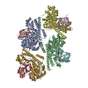
Mass: 384.411 Da / Num. of mol.: 1 / Source method: obtained synthetically / Formula: C14H20N6O5S / Feature type: SUBJECT OF INVESTIGATION
#4: Chemical
ChemComp-ZN / ZINC ION


Mass: 65.409 Da / Num. of mol.: 4 / Source method: obtained synthetically / Formula: Zn / Feature type: SUBJECT OF INVESTIGATION
#5: Water ChemComp-HOH / water


Mass: 18.015 Da / Num. of mol.: 1417 / Source method: isolated from a natural source / Formula: H2O
Has ligand of interestY

-
Experimental details

-
Experiment

ExperimentMethod: X-RAY DIFFRACTION / Number of used crystals: 1

-
Sample preparation

CrystalDensity Matthews: 2.37 Å3/Da / Density % sol: 48.04 %
Crystal growTemperature: 296 K / Method: vapor diffusion, hanging drop
Details: 0.02M Citric acid, 0.08M BIS-TRIS propane, pH8.8, 16% PEG 3350

-
Data collection

DiffractionMean temperature: 100 K / Serial crystal experiment: N
Diffraction sourceSource: SYNCHROTRON / Site: SSRF / Beamline: BL17B1 / Wavelength: 0.9785 Å
DetectorType: DECTRIS PILATUS3 S 6M / Detector: PIXEL / Date: Jul 3, 2020
RadiationProtocol: SINGLE WAVELENGTH / Monochromatic (M) / Laue (L): M / Scattering type: x-ray
Radiation wavelengthWavelength: 0.9785 Å / Relative weight: 1
Reflection twinOperator: h,-k,-h-l / Fraction: 0.09
ReflectionResolution: 3.5→49.34 Å / Num. obs: 76091 / % possible obs: 97.9 % / Redundancy: 3.51 % / CC1/2: 0.93 / Net I/σ(I): 2.29
Reflection shellResolution: 3.5→3.51 Å / Rmerge(I) obs: 2.09 / Num. unique obs: 11533

-
Processing

Software
NameVersionClassification
PHENIX1.19.2_4158refinement
PDB_EXTRACT3.27data extraction
XDSdata reduction
XDSdata scaling
MOLREPphasing
RefinementMethod to determine structure: MOLECULAR REPLACEMENT / Resolution: 3.52→19.99 Å / Cross valid method: THROUGHOUT / σ(F): 0 / Phase error: 31.23 / Stereochemistry target values: TWIN_LSQ_F
Details: There are 4 pairs of molecules in the ASU. Only chains A/C have been conducted in-depth analysis and optimization and used for further structure analysis. Other molecules have poor electron ...Details: There are 4 pairs of molecules in the ASU. Only chains A/C have been conducted in-depth analysis and optimization and used for further structure analysis. Other molecules have poor electron density and were not carried out in-depth optimization.
RfactorNum. reflection% reflection
Rfree0.2914 2021 5.11 %
Rwork0.2739 37546 -
obs0.2762 39534 99.34 %
Solvent computationShrinkage radii: 0.9 Å / VDW probe radii: 1.11 Å / Solvent model: FLAT BULK SOLVENT MODEL
Displacement parametersBiso max: 231.48 Å2 / Biso mean: 90.9712 Å2 / Biso min: 0.23 Å2
Refinement stepCycle: final / Resolution: 3.52→19.99 Å
ProteinNucleic acidLigandSolventTotal
Num. atoms19451 0 50 1526 21027
Biso mean--46.02 70.65 -
Num. residues----2474
LS refinement shell

Refine-ID: X-RAY DIFFRACTION / Rfactor Rfree error: 0 / Total num. of bins used: 14

Resolution (Å)Rfactor RfreeNum. reflection RfreeRfactor RworkNum. reflection RworkNum. reflection all% reflection obs (%)
3.52-3.60.32141420.27662612275492
3.6-3.70.33641390.28732613275295
3.7-3.810.24651370.25472707284495
3.81-3.930.26271460.24752627277395
3.93-4.070.26871370.26212691282895
4.07-4.230.26781390.25362662280195
4.23-4.420.27711380.24842725286395
4.42-4.650.28661430.2592616275994
4.65-4.940.29011380.26462639277793
4.94-5.310.2771460.27292722286895
5.31-5.840.30961470.30142693284095
5.84-6.650.31191410.30062688282995
6.66-8.280.30551500.28642773292395
8.28-19.990.31061450.29812778292394

+
About Yorodumi

-
News

-
Feb 9, 2022. New format data for meta-information of EMDB entries

New format data for meta-information of EMDB entries

  • Version 3 of the EMDB header file is now the official format.
  • The previous official version 1.9 will be removed from the archive.

Related info.:EMDB header

External links:wwPDB to switch to version 3 of the EMDB data model

-
Aug 12, 2020. Covid-19 info

Covid-19 info

URL: https://pdbjlc1.pdbj.org/emnavi/covid19.php

New page: Covid-19 featured information page in EM Navigator.

Related info.:Covid-19 info / Mar 5, 2020. Novel coronavirus structure data

+
Mar 5, 2020. Novel coronavirus structure data

Novel coronavirus structure data

Related info.:Yorodumi Speices / Aug 12, 2020. Covid-19 info

External links:COVID-19 featured content - PDBj / Molecule of the Month (242):Coronavirus Proteases

+
Jan 31, 2019. EMDB accession codes are about to change! (news from PDBe EMDB page)

EMDB accession codes are about to change! (news from PDBe EMDB page)

  • The allocation of 4 digits for EMDB accession codes will soon come to an end. Whilst these codes will remain in use, new EMDB accession codes will include an additional digit and will expand incrementally as the available range of codes is exhausted. The current 4-digit format prefixed with “EMD-” (i.e. EMD-XXXX) will advance to a 5-digit format (i.e. EMD-XXXXX), and so on. It is currently estimated that the 4-digit codes will be depleted around Spring 2019, at which point the 5-digit format will come into force.
  • The EM Navigator/Yorodumi systems omit the EMD- prefix.

Related info.:Q: What is EMD? / ID/Accession-code notation in Yorodumi/EM Navigator

External links:EMDB Accession Codes are Changing Soon! / Contact to PDBj

+
Jul 12, 2017. Major update of PDB

Major update of PDB

  • wwPDB released updated PDB data conforming to the new PDBx/mmCIF dictionary.
  • This is a major update changing the version number from 4 to 5, and with Remediation, in which all the entries are updated.
  • In this update, many items about electron microscopy experimental information are reorganized (e.g. em_software).
  • Now, EM Navigator and Yorodumi are based on the updated data.

External links:wwPDB Remediation / Enriched Model Files Conforming to OneDep Data Standards Now Available in the PDB FTP Archive

-
Yorodumi

Thousand views of thousand structures

  • Yorodumi is a browser for structure data from EMDB, PDB, SASBDB, etc.
  • This page is also the successor to EM Navigator detail page, and also detail information page/front-end page for Omokage search.
  • The word "yorodu" (or yorozu) is an old Japanese word meaning "ten thousand". "mi" (miru) is to see.

Related info.:EMDB / PDB / SASBDB / Comparison of 3 databanks / Yorodumi Search / Aug 31, 2016. New EM Navigator & Yorodumi / Yorodumi Papers / Jmol/JSmol / Function and homology information / Changes in new EM Navigator and Yorodumi

Read more