 Movie
Movie Controller
 Structure viewers
 About EMN search


-Search query
-Search result
Showing 1 - 50 of 245 items for (author: wagner & r)
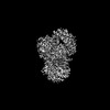
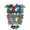
EMDB-42291: 
Structure of the human INTS9-INTS11-BRAT1 complex
EMDB-42292:
Structure of the Drosophila IntS11-CG7044(dBRAT1) complex

PDB-8uib:
Structure of the human INTS9-INTS11-BRAT1 complex

PDB-8uic:
Structure of the Drosophila IntS11-CG7044(dBRAT1) complex
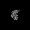
EMDB-18162:
N5-methyl-H4MPT:CoM methyltransferase -coenzyme M complex + CoM

EMDB-18135:
Cryo-EM structure of the methanogenic Na+ translocating N5-methyl-H4MPT:CoM methyltransferase complex
PDB-8q3v:
Cryo-EM structure of the methanogenic Na+ translocating N5-methyl-H4MPT:CoM methyltransferase complex

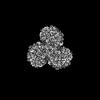
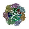
EMDB-17418:
CryoEM structure of a C7-symmetrical GroEL7-GroES7 cage in presence of ADP-BeFx

EMDB-17420:
CryoEM structure of a GroEL7-GroES7 cage with encapsulated disordered substrate MetK in the presence of ADP-BeFx

EMDB-17421:
CryoEM structure of a GroEL7-GroES7 cage with encapsulated ordered substrate MetK in the presence of ADP-BeFx
EMDB-17422:
Density for MetK encapsulated in the GroEL7-GroES7 cage

EMDB-17423:
Symmetry-averaged GroEL7-GroES7 chamber with encapsulated disordered substrate MetK obtained by in vitro cryo electron tomography

EMDB-17424:
Symmetry-averaged GroEL7-GroES7 chamber with encapsulated ordered substrate MetK obtained by in vitro cryo electron tomography
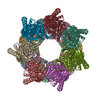
EMDB-17425:
Structure average of GroEL14 complexes found in the cytosol of Escherichia coli overexpressing GroEL obtained by cryo electron tomography

EMDB-17426:
In situ structure average of GroEL14-GroES14 complexes in Escherichia coli cytosol obtained by cryo electron tomography

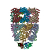
EMDB-17534:
Cryo-EM structure of a D7-symmetrical GroEL14-GroES14 complex in presence of ADP-BeFx

EMDB-17535:
Cryo-EM structure of a C7-symmetrical GroEL14-GroES7 complex in presence of ADP-BeFx

EMDB-17559:
In situ structure average of GroEL7-GroES7 chamber with no or disordered substrate in Escherichia coli cytosol obtained by cryo electron tomography

EMDB-17560:
In situ structure average of GroEL7-GroES7 chamber with encapsulated, ordered substrate in Escherichia coli cytosol obtained by cryo electron tomography

EMDB-17561:
Cryo-ET subtomogram of 70S ribosomes in Escherichia coli cells at 37 and 46 degrees centigrade and in Escherichia coli cells overexpressing GroELS and MetK

EMDB-17562:
Cryo-ET subtomogram of 70S ribosomes in Escherichia coli cells overexpressing GroEL

EMDB-17563:
CryoEM structure of a GroEL14-GroES7 cage with encapsulated ordered substrate MetK in the presence of ADP-BeFx

EMDB-17564:
CryoEM structure of a GroEL14-GroES7 cage with encapsulated disordered substrate MetK in the presence of ADP-BeFx

EMDB-17565:
CryoEM structure of a GroEL14-GroES14 cage with two encapsulated disordered MetK substrates in the presence of ADP-BeFx

EMDB-17566:
CryoEM structure of a (GroEL)14-(GroES)14 complex with encapsulated ordered MetK substrate in one chamber and no or disordered MetK substrate in the other chamber in the presence of ADP-BeFx

EMDB-17567:
Conformer 1 of the (GroEL)14(GroES)14 complex with two encapsulated, ordered and near-native MetK substrate molecules in the presence of ADP-BeFx

EMDB-17568:
Conformer 2 of the (GroEL)14(GroES)14 complex with two encapsulated, near-native and ordered MetK substrates in the presence of ADP-BeFx

EMDB-17569:
Conformer 3 of the (GroEL)14(GroES)14 complex with two encapsulated, near-native and ordered MetK substrates in the presence of ADP-BeFx

EMDB-17570:
Conformer 4 of the (GroEL)14(GroES)14 complex with two encapsulated, near-native and ordered MetK substrates in the presence of ADP-BeFx

EMDB-17571:
Conformer 5 of the (GroEL)14(GroES)14 complex with two encapsulated, near-native and ordered MetK substrates in the presence of ADP-BeFx

EMDB-17572:
Conformer 6 of the (GroEL)14(GroES)14 complex with two encapsulated, near-native and ordered MetK substrates in the presence of ADP-BeFx

EMDB-17573:
Conformer 7 of the (GroEL)14(GroES)14 complex with two encapsulated, near-native and ordered MetK substrates in the presence of ADP-BeFx

EMDB-18735:
CryoEM structure of a GroEL14-GroES7 complex in presence of ADP-BeFx with wide GroEL7 trans ring conformation

EMDB-18736:
CryoEM structure of a GroEL14-GroES7 complex in presence of ADP-BeFx with narrow GroEL7 trans ring conformation

EMDB-18737:
In situ structure average of GroEL14-GroES7 complexes with wide GroEL7 trans ring conformation in Escherichia coli cytosol obtained by cryo electron tomography

EMDB-18738:
In situ structure average of GroEL14-GroES7 complexes with narrow GroEL7 trans ring conformation in Escherichia coli cytosol obtained by cryo electron tomography
PDB-8p4m:
CryoEM structure of a C7-symmetrical GroEL7-GroES7 cage in presence of ADP-BeFx
PDB-8p4n:
CryoEM structure of a GroEL7-GroES7 cage with encapsulated disordered substrate MetK in the presence of ADP-BeFx
PDB-8p4o:
CryoEM structure of a GroEL7-GroES7 cage with encapsulated ordered substrate MetK in the presence of ADP-BeFx
PDB-8p4p:
Structure average of GroEL14 complexes found in the cytosol of Escherichia coli overexpressing GroEL obtained by cryo electron tomography

PDB-8p4r:
In situ structure average of GroEL14-GroES14 complexes in Escherichia coli cytosol obtained by cryo electron tomography
PDB-8qxs:
CryoEM structure of a GroEL14-GroES7 complex in presence of ADP-BeFx with wide GroEL7 trans ring conformation
PDB-8qxt:
CryoEM structure of a GroEL14-GroES7 complex in presence of ADP-BeFx with narrow GroEL7 trans ring conformation
PDB-8qxu:
In situ structure average of GroEL14-GroES7 complexes with wide GroEL7 trans ring conformation in Escherichia coli cytosol obtained by cryo electron tomography
PDB-8qxv:
In situ structure average of GroEL14-GroES7 complexes with narrow GroEL7 trans ring conformation in Escherichia coli cytosol obtained by cryo electron tomography

EMDB-16538:
Tomogram of Serratia marcescens in the post-spanin state (1)

EMDB-16539:
Tomogram of Serratia marcescens in the post-spanin state (2)

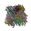
EMDB-16618:
Structure of Yersinia entomophaga Tc toxin from FIB-milled spheroplasts

EMDB-16619:
Tomogram of Yersinia entomophaga in the post-holin state (whole cell)

EMDB-16986:
Structure of the relaxed thin filament from FIB milled left ventricular mouse myofibrils (tropomyosin masked out)
Pages:
 wwPDB to switch to version 3 of the EMDB data model
