Movie
Movie Controller


+ Open data

- Basic information
| Entry |  | |||||||||
|---|---|---|---|---|---|---|---|---|---|---|
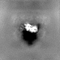
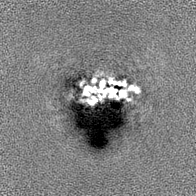
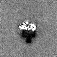
| Title | Density for MetK encapsulated in the GroEL7-GroES7 cage | |||||||||
 Map data Map data | Density for MetK encapsulated into the GroEL7-GroES7 cage | |||||||||
 Sample Sample |
| |||||||||
 Keywords Keywords | Chaperonin / Folding cage / proteostasis / heat shock / ATPase / CHAPERONE | |||||||||
| Function / homology |  Function and homology information Function and homology informationmethionine adenosyltransferase / methionine adenosyltransferase activity / S-adenosylmethionine biosynthetic process / L-methionine cycle / potassium ion binding / one-carbon metabolic process / magnesium ion binding / ATP binding / identical protein binding / cytosol Similarity search - Function | |||||||||
| Biological species |  | |||||||||
| Method | single particle reconstruction / cryo EM / Resolution: 3.7 Å | |||||||||
 Authors Authors | Wagner J / Caravajal AI / Beck F / Bracher A / Wan W / Bohn S / Koerner R / Fernandez-Busnadiego R / Baumeister W / Hartl FU | |||||||||
| Funding support | 1 items
| |||||||||
 Citation Citation | Journal: Nature / Year: 2024 Title: Visualizing chaperonin function in situ by cryo-electron tomography. Authors: Jonathan Wagner / Alonso I Carvajal / Andreas Bracher / Florian Beck / William Wan / Stefan Bohn / Roman Körner / Wolfgang Baumeister / Ruben Fernandez-Busnadiego / F Ulrich Hartl /   Abstract: Chaperonins are large barrel-shaped complexes that mediate ATP-dependent protein folding. The bacterial chaperonin GroEL forms juxtaposed rings that bind unfolded protein and the lid-shaped cofactor ...Chaperonins are large barrel-shaped complexes that mediate ATP-dependent protein folding. The bacterial chaperonin GroEL forms juxtaposed rings that bind unfolded protein and the lid-shaped cofactor GroES at their apertures. In vitro analyses of the chaperonin reaction have shown that substrate protein folds, unimpaired by aggregation, while transiently encapsulated in the GroEL central cavity by GroES. To determine the functional stoichiometry of GroEL, GroES and client protein in situ, here we visualized chaperonin complexes in their natural cellular environment using cryo-electron tomography. We find that, under various growth conditions, around 55-70% of GroEL binds GroES asymmetrically on one ring, with the remainder populating symmetrical complexes. Bound substrate protein is detected on the free ring of the asymmetrical complex, defining the substrate acceptor state. In situ analysis of GroEL-GroES chambers, validated by high-resolution structures obtained in vitro, showed the presence of encapsulated substrate protein in a folded state before release into the cytosol. Based on a comprehensive quantification and conformational analysis of chaperonin complexes, we propose a GroEL-GroES reaction cycle that consists of linked asymmetrical and symmetrical subreactions mediating protein folding. Our findings illuminate the native conformational and functional chaperonin cycle directly within cells. | |||||||||
| History |
|
- Structure visualization
| Supplemental images |
|---|

- Downloads & links
-EMDB archive
| Map data | emd_17422.map.gz | 27 MB | EMDB map data format | |
|---|---|---|---|---|
| Header (meta data) | emd-17422-v30.xmlemd-17422.xml | 21.3 KB 21.3 KB | Display Display | EMDB header |
| FSC (resolution estimation) | emd_17422_fsc.xml | 6.4 KB | Display | FSC data file |
| Images |  emd_17422.png emd_17422.png | 55.9 KB | ||
| Filedesc metadata | emd-17422.cif.gz | 5.6 KB | ||
| Others | emd_17422_half_map_1.map.gzemd_17422_half_map_2.map.gz | 26.7 MB 26.7 MB | ||
| Archive directory |  http://ftp.pdbj.org/pub/emdb/structures/EMD-17422ftp://ftp.pdbj.org/pub/emdb/structures/EMD-17422 http://ftp.pdbj.org/pub/emdb/structures/EMD-17422ftp://ftp.pdbj.org/pub/emdb/structures/EMD-17422 | HTTPS FTP |
-Related structure data
| Related structure data | 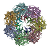 8p4mC 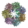 8p4nC 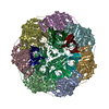 8p4oC 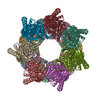 8p4pC  8p4rC 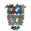 8qxsC 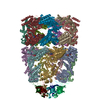 8qxtC 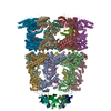 8qxuC 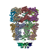 8qxvC C: citing same article ( |
|---|---|
| Similar structure data |


-Links
| EMDB pages | EMDB (EBI/PDBe) / EMDataResource |
|---|
-Map
| File | Download / File: emd_17422.map.gz / Format: CCP4 / Size: 28.7 MB / Type: IMAGE STORED AS FLOATING POINT NUMBER (4 BYTES) | ||||||||||||||||||||||||||||||||||||
|---|---|---|---|---|---|---|---|---|---|---|---|---|---|---|---|---|---|---|---|---|---|---|---|---|---|---|---|---|---|---|---|---|---|---|---|---|---|
| Annotation | Density for MetK encapsulated into the GroEL7-GroES7 cage | ||||||||||||||||||||||||||||||||||||
| Projections & slices | Image control
Images are generated by Spider. | ||||||||||||||||||||||||||||||||||||
| Voxel size | X=Y=Z: 1.09 Å | ||||||||||||||||||||||||||||||||||||
| Density |
| ||||||||||||||||||||||||||||||||||||
| Symmetry | Space group: 1 | ||||||||||||||||||||||||||||||||||||
| Details | EMDB XML:
|
 Z (Sec.)
 Y (Row.)
 X (Col.)


-Supplemental data
-Half map: #1
| File | emd_17422_half_map_1.map | ||||||||||||
|---|---|---|---|---|---|---|---|---|---|---|---|---|---|
| Projections & Slices |
| ||||||||||||
| Density Histograms |


- Sample components
-Entire : GroEL14-GroES14-MetK2
| Entire | Name: GroEL14-GroES14-MetK2 |
|---|---|
| Components |
|
-Supramolecule #1: GroEL14-GroES14-MetK2
| Supramolecule | Name: GroEL14-GroES14-MetK2 / type: complex / ID: 1 / Parent: 0 / Macromolecule list: all / Details: with bound ADP-BeF3 Mg2+ K+ |
|---|---|
| Source (natural) | Organism: |
| Molecular weight | Theoretical: 1.03 MDa |
-Macromolecule #1: MetK
| Macromolecule | Name: MetK / type: protein_or_peptide / ID: 1 / Enantiomer: LEVO / EC number: methionine adenosyltransferase |
|---|---|
| Source (natural) | Organism: |
| Recombinant expression | Organism: |
| Sequence | String: MAKHLFTSES VSEGHPDKIA DQISDAVLDA ILEQDPKARV ACETYVKTGM VLVGGEITTS AWVDIEEIT RNTVREIGYV HSDMGFDANS CAVLSAIGKQ SPDINQGVDR ADPLEQGAGD Q GLMFGYAT NETDVLMPAP ITYAHRLVQR QAEVRKNGTL PWLRPDAKSQ ...String: MAKHLFTSES VSEGHPDKIA DQISDAVLDA ILEQDPKARV ACETYVKTGM VLVGGEITTS AWVDIEEIT RNTVREIGYV HSDMGFDANS CAVLSAIGKQ SPDINQGVDR ADPLEQGAGD Q GLMFGYAT NETDVLMPAP ITYAHRLVQR QAEVRKNGTL PWLRPDAKSQ VTFQYDDGKI VG IDAVVLS TQHSEEIDQK SLQEAVMEEI IKPILPAEWL TSATKFFINP TGRFVIGGPM GDC GLTGRK IIVDTYGGMA RHGGGAFSGK DPSKVDRSAA YAARYVAKNI VAAGLADRCE IQVS YAIGV AEPTSIMVET FGTEKVPSEQ LTLLVREFFD LRPYGLIQML DLLHPIYKET AAYGH FGRE HFPWEKTDKA QLLRDAAGLK UniProtKB: S-adenosylmethionine synthase |
-Experimental details
-Structure determination
| Method | cryo EM |
|---|---|
 Processing Processing | single particle reconstruction |
| Aggregation state | particle |
-Sample preparation
| Concentration | 10.3 mg/mL | ||||||||||||||||||||||||
|---|---|---|---|---|---|---|---|---|---|---|---|---|---|---|---|---|---|---|---|---|---|---|---|---|---|
| Buffer | pH: 7.4 Component:
| ||||||||||||||||||||||||
| Grid | Model: Quantifoil R2/1 / Material: COPPER / Mesh: 200 / Support film - Material: CARBON / Support film - topology: HOLEY / Pretreatment - Type: PLASMA CLEANING / Pretreatment - Time: 30 sec. | ||||||||||||||||||||||||
| Vitrification | Cryogen name: ETHANE-PROPANE / Chamber humidity: 100 % / Chamber temperature: 277 K / Instrument: FEI VITROBOT MARK IV Details: 5.9 mM n-octyl-beta-D-glucopyranoside were added before vitrification. |
- Electron microscopy
| Microscope | FEI TITAN KRIOS |
|---|---|
| Image recording | Film or detector model: GATAN K3 (6k x 4k) / Average electron dose: 55.0 e/Å2 |
| Electron beam | Acceleration voltage: 300 kV / Electron source:  FIELD EMISSION GUN FIELD EMISSION GUN |
| Electron optics | Illumination mode: OTHER / Imaging mode: BRIGHT FIELD / Nominal defocus max: 3.0 µm / Nominal defocus min: 0.5 µm / Nominal magnification: 22500 |
| Experimental equipment |  Model: Titan Krios / Image courtesy: FEI Company |