 Movie
Movie Controller

[English] 日本語
 Yorodumi
- EMDB-17559: In situ structure average of GroEL7-GroES7 chamber with no or dis... -
+ Open data

- Basic information
| Entry |  | |||||||||
|---|---|---|---|---|---|---|---|---|---|---|
| Title | In situ structure average of GroEL7-GroES7 chamber with no or disordered substrate in Escherichia coli cytosol obtained by cryo electron tomography | |||||||||
 Map data Map data | ||||||||||
 Sample Sample |
| |||||||||
 Keywords Keywords | Chaperonin / Folding cage / proteostasis / heat shock / ATPase / CHAPERONE | |||||||||
| Function / homology |  Function and homology information Function and homology informationGroEL-GroES complex / chaperonin ATPase / virion assembly / isomerase activity / protein folding chaperone / ATP-dependent protein folding chaperone / response to radiation / : / protein refolding / response to heat ...GroEL-GroES complex / chaperonin ATPase / virion assembly / isomerase activity / protein folding chaperone / ATP-dependent protein folding chaperone / response to radiation / : / protein refolding / response to heat / protein-folding chaperone binding / protein folding / magnesium ion binding / ATP hydrolysis activity / ATP binding / membrane / metal ion binding / identical protein binding / cytosol Similarity search - Function | |||||||||
| Biological species |  | |||||||||
| Method | subtomogram averaging / cryo EM / Resolution: 15.1 Å | |||||||||
 Authors Authors | Wagner J / Caravajal AI / Beck F / Bracher A / Wan W / Bohn S / Koerner R / Baumeister W / Fernandez-Busnadiego R / Hartl FU | |||||||||
| Funding support | 1 items
| |||||||||
 Citation Citation | Journal: Nature / Year: 2024 Title: Visualizing chaperonin function in situ by cryo-electron tomography. Authors: Jonathan Wagner / Alonso I Carvajal / Andreas Bracher / Florian Beck / William Wan / Stefan Bohn / Roman Körner / Wolfgang Baumeister / Ruben Fernandez-Busnadiego / F Ulrich Hartl /   Abstract: Chaperonins are large barrel-shaped complexes that mediate ATP-dependent protein folding. The bacterial chaperonin GroEL forms juxtaposed rings that bind unfolded protein and the lid-shaped cofactor ...Chaperonins are large barrel-shaped complexes that mediate ATP-dependent protein folding. The bacterial chaperonin GroEL forms juxtaposed rings that bind unfolded protein and the lid-shaped cofactor GroES at their apertures. In vitro analyses of the chaperonin reaction have shown that substrate protein folds, unimpaired by aggregation, while transiently encapsulated in the GroEL central cavity by GroES. To determine the functional stoichiometry of GroEL, GroES and client protein in situ, here we visualized chaperonin complexes in their natural cellular environment using cryo-electron tomography. We find that, under various growth conditions, around 55-70% of GroEL binds GroES asymmetrically on one ring, with the remainder populating symmetrical complexes. Bound substrate protein is detected on the free ring of the asymmetrical complex, defining the substrate acceptor state. In situ analysis of GroEL-GroES chambers, validated by high-resolution structures obtained in vitro, showed the presence of encapsulated substrate protein in a folded state before release into the cytosol. Based on a comprehensive quantification and conformational analysis of chaperonin complexes, we propose a GroEL-GroES reaction cycle that consists of linked asymmetrical and symmetrical subreactions mediating protein folding. Our findings illuminate the native conformational and functional chaperonin cycle directly within cells. | |||||||||
| History |
|
- Structure visualization
| Supplemental images |
|---|

- Downloads & links
-EMDB archive
| Map data | emd_17559.map.gz | 7.3 MB | EMDB map data format | |
|---|---|---|---|---|
| Header (meta data) | emd-17559-v30.xmlemd-17559.xml | 16.2 KB 16.2 KB | Display Display | EMDB header |
| FSC (resolution estimation) | emd_17559_fsc.xml | 4.4 KB | Display | FSC data file |
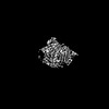
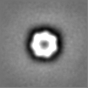
| Images |  emd_17559.png emd_17559.png | 23.7 KB | ||
| Masks | emd_17559_msk_1.map | 8 MB | Mask map | |
| Filedesc metadata | emd-17559.cif.gz | 5.3 KB | ||
| Others | emd_17559_half_map_1.map.gzemd_17559_half_map_2.map.gz | 7.4 MB 7.4 MB | ||
| Archive directory |  http://ftp.pdbj.org/pub/emdb/structures/EMD-17559ftp://ftp.pdbj.org/pub/emdb/structures/EMD-17559 http://ftp.pdbj.org/pub/emdb/structures/EMD-17559ftp://ftp.pdbj.org/pub/emdb/structures/EMD-17559 | HTTPS FTP |
-Related structure data
| Related structure data | 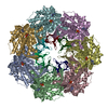 8p4mC 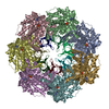 8p4nC 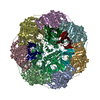 8p4oC 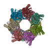 8p4pC  8p4rC  8qxsC  8qxtC  8qxuC  8qxvC  17427 C: citing same article ( |
|---|---|
| Similar structure data |


-Links
| EMDB pages | EMDB (EBI/PDBe) / EMDataResource |
|---|---|
| Related items in Molecule of the Month |


-Map
| File | Download / File: emd_17559.map.gz / Format: CCP4 / Size: 8 MB / Type: IMAGE STORED AS FLOATING POINT NUMBER (4 BYTES) | ||||||||||||||||||||||||||||||||||||
|---|---|---|---|---|---|---|---|---|---|---|---|---|---|---|---|---|---|---|---|---|---|---|---|---|---|---|---|---|---|---|---|---|---|---|---|---|---|
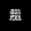
| Projections & slices | Image control
Images are generated by Spider. | ||||||||||||||||||||||||||||||||||||
| Voxel size | X=Y=Z: 3.52 Å | ||||||||||||||||||||||||||||||||||||
| Density |
| ||||||||||||||||||||||||||||||||||||
| Symmetry | Space group: 1 | ||||||||||||||||||||||||||||||||||||
| Details | EMDB XML:
|
 Z (Sec.)
 Y (Row.)
 X (Col.)


-Supplemental data
-Mask #1
| File | emd_17559_msk_1.map | ||||||||||||
|---|---|---|---|---|---|---|---|---|---|---|---|---|---|
| Projections & Slices |
| ||||||||||||
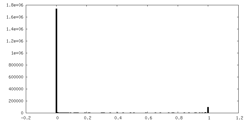
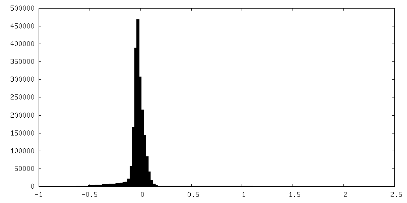
| Density Histograms |


-Half map: #1
| File | emd_17559_half_map_1.map | ||||||||||||
|---|---|---|---|---|---|---|---|---|---|---|---|---|---|
| Projections & Slices |
| ||||||||||||
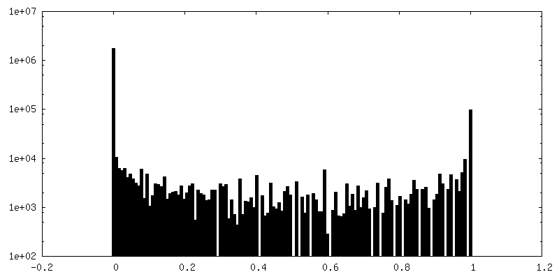
| Density Histograms |


- Sample components
-Entire : GroEL7-GroES7 subcomplex
| Entire | Name: GroEL7-GroES7 subcomplex |
|---|---|
| Components |
|
-Supramolecule #1: GroEL7-GroES7 subcomplex
| Supramolecule | Name: GroEL7-GroES7 subcomplex / type: complex / ID: 1 / Parent: 0 / Macromolecule list: all Details: subtomograms of GroEL14-GroES14 and GroEL14-GroES7 complexes in E. coli cytosol were aligned so that GroEL7-GroES7 subcomplexes with density for no or disordered substrate match |
|---|---|
| Source (natural) | Organism: |
-Macromolecule #1: GroEL
| Macromolecule | Name: GroEL / type: protein_or_peptide / ID: 1 / Enantiomer: LEVO |
|---|---|
| Source (natural) | Organism: |
| Sequence | String: AAKDVKFGND ARVKMLRGVN VLADAVKVTL GPKGRNVVLD KSFGAPTITK DGVSVAREI ELEDKFENMG AQMVKEVASK ANDAAGDGTT TATVLAQAII TEGLKAVAAG MNPMDLKRGI DKAVTAAVE ELKALSVPCS DSKAIAQVGT ISANSDETVG KLIAEAMDKV ...String: AAKDVKFGND ARVKMLRGVN VLADAVKVTL GPKGRNVVLD KSFGAPTITK DGVSVAREI ELEDKFENMG AQMVKEVASK ANDAAGDGTT TATVLAQAII TEGLKAVAAG MNPMDLKRGI DKAVTAAVE ELKALSVPCS DSKAIAQVGT ISANSDETVG KLIAEAMDKV GKEGVITVED G TGLQDELD VVEGMQFDRG YLSPYFINKP ETGAVELESP FILLADKKIS NIREMLPVLE AV AKAGKPL LIIAEDVEGE ALATLVVNTM RGIVKVAAVK APGFGDRRKA MLQDIATLTG GTV ISEEIG MELEKATLED LGQAKRVVIN KDTTTIIDGV GEEAAIQGRV AQIRQQIEEA TSDY DREKL QERVAKLAGG VAVIKVGAAT EVEMKEKKAR VEDALHATRA AVEEGVVAGG GVALI RVAS KLADLRGQNE DQNVGIKVAL RAMEAPLRQI VLNCGEEPSV VANTVKGGDG NYGYNA ATE EYGNMIDMGI LDPTKVTRSA LQYAASVAGL MITTECMVTD LPKNDAADLG AAGGMGG MG GMGGMM UniProtKB: Chaperonin GroEL |
-Macromolecule #2: GroES
| Macromolecule | Name: GroES / type: protein_or_peptide / ID: 2 / Enantiomer: LEVO |
|---|---|
| Source (natural) | Organism: |
| Sequence | String: MNIRPLHDRV IVKRKEVETK SAGGIVLTGS AAAKSTRGEV LAVGNGRILE NGEVKPLDVK VGDIVIFND GYGVKSEKID NEEVLIMSES DILAIVEA UniProtKB: Co-chaperonin GroES |
-Experimental details
-Structure determination
| Method | cryo EM |
|---|---|
 Processing Processing | subtomogram averaging |
| Aggregation state | cell |
-Sample preparation
| Buffer | pH: 7.4 |
|---|---|
| Vitrification | Cryogen name: ETHANE-PROPANE |
| Details | Vitrified E. coli Bl21 (DE3) cells |
- Electron microscopy
| Microscope | FEI TITAN KRIOS |
|---|---|
| Image recording | Film or detector model: GATAN K2 SUMMIT (4k x 4k) / Detector mode: SUPER-RESOLUTION / Average electron dose: 3.0 e/Å2 |
| Electron beam | Acceleration voltage: 300 kV / Electron source:  FIELD EMISSION GUN FIELD EMISSION GUN |
| Electron optics | C2 aperture diameter: 70.0 µm / Illumination mode: FLOOD BEAM / Imaging mode: BRIGHT FIELD / Cs: 2.7 mm / Nominal defocus max: 5.0 µm / Nominal defocus min: 2.5 µm |
| Experimental equipment |  Model: Titan Krios / Image courtesy: FEI Company |
-Image processing
| Final reconstruction | Applied symmetry - Point group: C1 (asymmetric) / Resolution.type: BY AUTHOR / Resolution: 15.1 Å / Resolution method: FSC 0.143 CUT-OFF / Software - Name: STOPGAP / Number subtomograms used: 24435 |
|---|---|
| Extraction | Number tomograms: 216 / Number images used: 125860 |
| Final angle assignment | Type: MAXIMUM LIKELIHOOD / Software - Name: RELION |
| FSC plot (resolution estimation) | 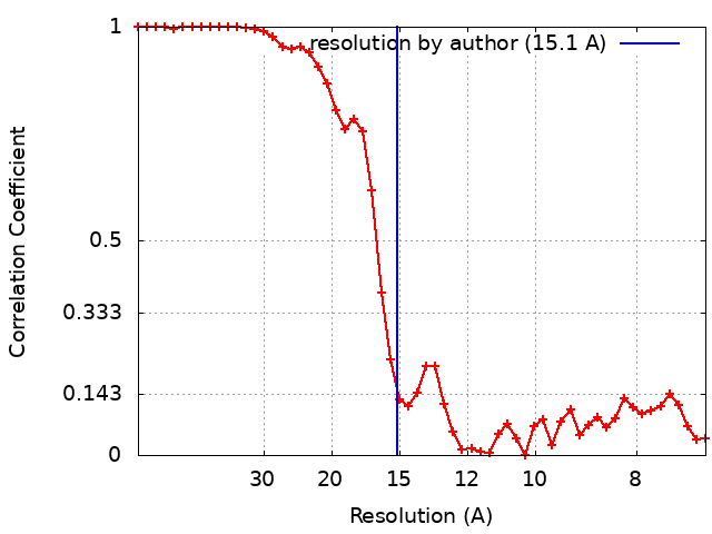 |
