 Movie
Movie Controller
 Structure viewers
 About EMN search


-Search query
-Search result
Showing 1 - 50 of 53 items for (author: righetto & r)

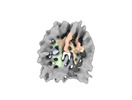
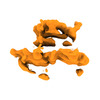
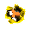
EMDB-41105: 
CryoEM structure of human DDB1-DCAF12 in complex with MAGEA3

PDB-8t9a:
CryoEM structure of human DDB1-DCAF12 in complex with MAGEA3
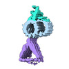
EMDB-19906:
In situ nucleosome subtomogram average from Chlamydomonas reinhardtii

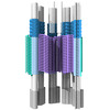
EMDB-16451:
Subtomogram average of the T. kivui 70S ribosome in situ

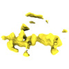
EMDB-15252:
In situ subtomogram average of the C. reinhardtii stellate at the ciliary transition zone

EMDB-15253:
In situ subtomogram average of the C. reinhardtii Y-link at the ciliary transition zone

EMDB-15254:
In situ subtomogram average of the C. reinhardtii MTD sleeve at the ciliary transition zone

EMDB-15255:
In situ subtomogram average of the C. reinhardtii MTD at the ciliary transition zone
EMDB-15256:
Composite map of the C. reinhardtii ciliary transition zone (structures attached to a single MTD) from in situ subtomogram averaging
EMDB-15257:
Composite map of the C. reinhardtii ciliary transition zone (full 9-fold assembly) from in situ subtomogram averaging
EMDB-15258:
In situ subtomogram average of C. reinhardtii IFT-B at the ciliary transition zone
EMDB-15259:
In situ subtomogram average of C. reinhardtii IFT-A at the ciliary transition zone

EMDB-15260:
In situ subtomogram average of C. reinhardtii dynein-1b at the ciliary transition zone
EMDB-15261:
Composite map of the C. reinhardtii IFT (segment of a fully assembled train) at the ciliary transition zone
EMDB-15262:
Tomogram #3 of the C. reinhardtii ciliary transition zone

EMDB-15263:
Tomogram #12 of the C. reinhardtii ciliary transition zone
EMDB-14169:
Cryo-EM structure of Hydrogen-dependent CO2 reductase.
EMDB-15053:
In situ structure of HDCR filaments

EMDB-15054:
In situ structure of the T. kivui ribosome

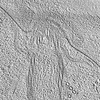
EMDB-15055:
In situ cryo-electron tomogram of a T. kivui cell 1

EMDB-15056:
In situ cryo-electron tomogram of a T. kivui cell 2
PDB-7qv7:
Cryo-EM structure of Hydrogen-dependent CO2 reductase.
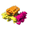
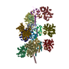
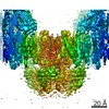
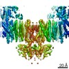
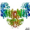
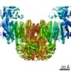
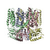
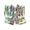
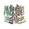
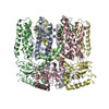
EMDB-12073:
Cryo-EM Structure of Human Thyroglobulin

PDB-7b75:
Cryo-EM Structure of Human Thyroglobulin

EMDB-10615:
FAK structure from single particle analysis of 2D crystals

EMDB-10616:
FAK structure with AMP-PNP from single particle analysis of 2D crystals

PDB-6ty3:
FAK structure from single particle analysis of 2D crystals

PDB-6ty4:
FAK structure with AMP-PNP from single particle analysis of 2D crystals

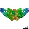
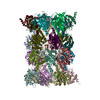
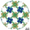
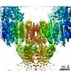
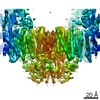
EMDB-10835:
High resolution cryo-EM structure of urease from the pathogen Yersinia enterocolitica

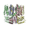
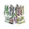
PDB-6yl3:
High resolution cryo-EM structure of urease from the pathogen Yersinia enterocolitica
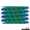
EMDB-4628:
Cryo-EM structure of Tobacco Mossaic Virus from microfluidic grid preparation.

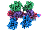
EMDB-4738:
Endogeneous native human 20S proteasome with bound Fabs isolated from less than 1 ul cell lysate
PDB-6r70:
Endogeneous native human 20S proteasome

PDB-6r7m:
Tobacco Mosaic Virus (TMV)
EMDB-4432:
MloK1 consensus map from single particle analysis of 2D crystals
EMDB-4439:
Full MloK1 consensus map from single particle analysis of 2D crystals
EMDB-4441:
MloK1 map from single particle analysis of 2D crystals, class 1 (extended conformation)
EMDB-4513:
MloK1 map from single particle analysis of 2D crystals, class 2 (intermediate conformation)
EMDB-4514:
MloK1 map from single particle analysis of 2D crystals, class 3 (intermediate extended conformation)
EMDB-4515:
MloK1 map from single particle analysis of 2D crystals, class 4 (compact/open conformation)
EMDB-4516:
MloK1 map from single particle analysis of 2D crystals, class 5 (intermediate compact conformation)

EMDB-4517:
MloK1 map from single particle analysis of 2D crystals, class 6 (intermediate compact conformation)
EMDB-4518:
MloK1 map from single particle analysis of 2D crystals, class 7 (intermediate conformation)
EMDB-4519:
MloK1 model from single particle analysis of 2D crystals, class 8 (intermediate conformation)
PDB-6i9d:
MloK1 consensus structure from single particle analysis of 2D crystals
PDB-6iax:
MloK1 model from single particle analysis of 2D crystals, class 1 (extended conformation)
PDB-6qcy:
MloK1 model from single particle analysis of 2D crystals, class 2 (intermediate conformation)
PDB-6qcz:
MloK1 model from single particle analysis of 2D crystals, class 3 (intermediate extended conformation)
PDB-6qd0:
MloK1 model from single particle analysis of 2D crystals, class 4 (compact/open conformation)
PDB-6qd1:
MloK1 model from single particle analysis of 2D crystals, class 5 (intermediate compact conformation)
Pages:
 wwPDB to switch to version 3 of the EMDB data model
