 Movie
Movie Controller

[English] 日本語
 Yorodumi
- PDB-6yl3: High resolution cryo-EM structure of urease from the pathogen Yer... -
+ Open data

- Basic information
| Entry | Database: PDB / ID: 6yl3 | |||||||||||||||||||||||||||||||||||||||||||||
|---|---|---|---|---|---|---|---|---|---|---|---|---|---|---|---|---|---|---|---|---|---|---|---|---|---|---|---|---|---|---|---|---|---|---|---|---|---|---|---|---|---|---|---|---|---|---|
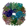
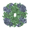
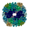
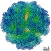
| Title | High resolution cryo-EM structure of urease from the pathogen Yersinia enterocolitica | |||||||||||||||||||||||||||||||||||||||||||||
 Components Components |
| |||||||||||||||||||||||||||||||||||||||||||||
 Keywords Keywords | METAL BINDING PROTEIN / Urease / enzyme / nickel / metalloenzyme / pathogen | |||||||||||||||||||||||||||||||||||||||||||||
| Function / homology |  Function and homology information Function and homology informationurease complex / urease / urease activity / urea catabolic process / nickel cation binding / cytoplasm Similarity search - Function | |||||||||||||||||||||||||||||||||||||||||||||
| Biological species |  Yersinia enterocolitica W22703 (bacteria) Yersinia enterocolitica W22703 (bacteria) | |||||||||||||||||||||||||||||||||||||||||||||
| Method | ELECTRON MICROSCOPY / single particle reconstruction / cryo EM / Resolution: 1.98 Å | |||||||||||||||||||||||||||||||||||||||||||||
 Authors Authors | Righetto, R.D. / Anton, L. / Adaixo, R. / Jakob, R. / Zivanov, J. / Mahi, M.A. / Ringler, P. / Schwede, T. / Maier, T. / Stahlberg, H. | |||||||||||||||||||||||||||||||||||||||||||||
| Funding support |  Switzerland, 1items Switzerland, 1items
| |||||||||||||||||||||||||||||||||||||||||||||
 Citation Citation | Journal: Nat Commun / Year: 2020 Title: High-resolution cryo-EM structure of urease from the pathogen Yersinia enterocolitica. Authors: Ricardo D Righetto / Leonie Anton / Ricardo Adaixo / Roman P Jakob / Jasenko Zivanov / Mohamed-Ali Mahi / Philippe Ringler / Torsten Schwede / Timm Maier / Henning Stahlberg / Abstract: Urease converts urea into ammonia and carbon dioxide and makes urea available as a nitrogen source for all forms of life except animals. In human bacterial pathogens, ureases also aid in the invasion ...Urease converts urea into ammonia and carbon dioxide and makes urea available as a nitrogen source for all forms of life except animals. In human bacterial pathogens, ureases also aid in the invasion of acidic environments such as the stomach by raising the surrounding pH. Here, we report the structure of urease from the pathogen Yersinia enterocolitica at 2 Å resolution from cryo-electron microscopy. Y. enterocolitica urease is a dodecameric assembly of a trimer of three protein chains, ureA, ureB and ureC. The high data quality enables detailed visualization of the urease bimetal active site and of the impact of radiation damage. The obtained structure is of sufficient quality to support drug development efforts. #1: Journal: Biorxiv / Year: 2020Title: High-resolution cryo-EM structure of urease from the pathogen Yersinia enterocolitica Authors: Righetto, R.D. / Anton, L. / Adaixo, R. / Jakob, R. / Zivanov, J. / Mahi, M.A. / Ringler, P. / Schwede, T. / Maier, T. / Stahlberg, H. | |||||||||||||||||||||||||||||||||||||||||||||
| History |
|
- Structure visualization
| Movie |
Movie viewer |
|---|---|
| Structure viewer | Molecule: MolmilJmol/JSmol |
- Downloads & links
-Download
| PDBx/mmCIF format | 6yl3.cif.gz | 3 MB | Display | PDBx/mmCIF format |
|---|---|---|---|---|
| PDB format | pdb6yl3.ent.gz | Display | PDB format | |
| PDBx/mmJSON format | 6yl3.json.gz | Tree view | PDBx/mmJSON format | |
| Others |  Other downloads Other downloads |
-Validation report
| Arichive directory | https://data.pdbj.org/pub/pdb/validation_reports/yl/6yl3ftp://data.pdbj.org/pub/pdb/validation_reports/yl/6yl3 | HTTPS FTP |
|---|
-Related structure data
| Related structure data |  10835MC M: map data used to model this data C: citing same article ( |
|---|---|
| Similar structure data | |
| EM raw data | EMPIAR-10389 (Title: High resolution cryo-EM structure of urease from the pathogen Yersinia enterocolitica Data size: 839.9 Data #1: Aligned averages of Y. enterocolitica urease [micrographs - single frame] Data #2: Raw multi-frame micrographs of Y. enterocolitica urease [micrographs - multiframe]) |


-Links
 PDBj
PDBj

- Assembly
| Deposited unit | 
|
|---|---|
| 1 |
|
-Components
| #1: Protein | Mass: 11063.837 Da / Num. of mol.: 12 / Source method: isolated from a natural source / Source: (natural) Yersinia enterocolitica W22703 (bacteria) / References: UniProt: F4MWM9, urease#2: Protein | Mass: 14611.317 Da / Num. of mol.: 12 / Source method: isolated from a natural source / Source: (natural) Yersinia enterocolitica W22703 (bacteria) / References: UniProt: F4MWM8, urease#3: Protein | Mass: 61054.859 Da / Num. of mol.: 12 / Source method: isolated from a natural source / Source: (natural) Yersinia enterocolitica W22703 (bacteria) / References: UniProt: F4MWM7, urease#4: Chemical | ChemComp-NI /   Mass: 58.693 Da / Num. of mol.: 24 / Source method: obtained synthetically / Formula: Ni Mass: 58.693 Da / Num. of mol.: 24 / Source method: obtained synthetically / Formula: Ni#5: Water | ChemComp-HOH / |  Mass: 18.015 Da / Num. of mol.: 3672 / Source method: isolated from a natural source / Formula: H2O Mass: 18.015 Da / Num. of mol.: 3672 / Source method: isolated from a natural source / Formula: H2OHas ligand of interest | N | Has protein modification | Y | |
|---|
-Experimental details
-Experiment
| Experiment | Method: ELECTRON MICROSCOPY |
|---|---|
| EM experiment | Aggregation state: PARTICLE / 3D reconstruction method: single particle reconstruction |
- Sample preparation
| Component | Name: Urease oligomer from Y. enterocolitica / Type: COMPLEX / Entity ID: #1-#3 / Source: NATURAL |
|---|---|
| Molecular weight | Value: 1.025 MDa / Experimental value: YES |
| Source (natural) | Organism: Yersinia enterocolitica W22703 (bacteria) / Strain: E40 |
| Buffer solution | pH: 7 |
| Specimen | Conc.: 0.39 mg/ml / Embedding applied: NO / Shadowing applied: NO / Staining applied: NO / Vitrification applied: YES |
| Specimen support | Grid material: COPPER / Grid mesh size: 200 divisions/in. / Grid type: Quantifoil R1.2/1.3 |
| Vitrification | Instrument: FEI VITROBOT MARK IV / Cryogen name: ETHANE / Humidity: 90 % / Chamber temperature: 293.15 K / Details: 3 seconds blotting |
- Electron microscopy imaging
| Experimental equipment |  Model: Titan Krios / Image courtesy: FEI Company |
|---|---|
| Microscopy | Model: FEI TITAN KRIOS |
| Electron gun | Electron source:  FIELD EMISSION GUN / Accelerating voltage: 300 kV / Illumination mode: FLOOD BEAM FIELD EMISSION GUN / Accelerating voltage: 300 kV / Illumination mode: FLOOD BEAM |
| Electron lens | Mode: BRIGHT FIELD / Cs: 2.7 mm / C2 aperture diameter: 70 µm |
| Specimen holder | Cryogen: NITROGEN / Specimen holder model: FEI TITAN KRIOS AUTOGRID HOLDER |
| Image recording | Electron dose: 42 e/Å2 / Detector mode: COUNTING / Film or detector model: GATAN K2 SUMMIT (4k x 4k) |
| EM imaging optics | Energyfilter name: GIF Quantum LS / Energyfilter slit width: 20 eV |
- Processing
| EM software |
| ||||||||||||||||||||||||||||
|---|---|---|---|---|---|---|---|---|---|---|---|---|---|---|---|---|---|---|---|---|---|---|---|---|---|---|---|---|---|
| CTF correction | Type: PHASE FLIPPING AND AMPLITUDE CORRECTION | ||||||||||||||||||||||||||||
| Symmetry | Point symmetry: T (tetrahedral) | ||||||||||||||||||||||||||||
| 3D reconstruction | Resolution: 1.98 Å / Resolution method: FSC 0.143 CUT-OFF / Num. of particles: 97627 / Algorithm: FOURIER SPACE / Symmetry type: POINT |
