Movie
Movie Controller

[English] 日本語
 Yorodumi
- PDB-6zja: Helicobacter pylori urease with inhibitor bound in the active site -
+ Open data

- Basic information
| Entry | Database: PDB / ID: 6zja | |||||||||||||||||||||||||||||||||||||||||||||||||||
|---|---|---|---|---|---|---|---|---|---|---|---|---|---|---|---|---|---|---|---|---|---|---|---|---|---|---|---|---|---|---|---|---|---|---|---|---|---|---|---|---|---|---|---|---|---|---|---|---|---|---|---|---|
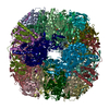
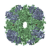
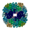
| Title | Helicobacter pylori urease with inhibitor bound in the active site | |||||||||||||||||||||||||||||||||||||||||||||||||||
 Components Components |
| |||||||||||||||||||||||||||||||||||||||||||||||||||
 Keywords Keywords | HYDROLASE / dodecamer / bi nickel center / enzyme / cytoplasm | |||||||||||||||||||||||||||||||||||||||||||||||||||
| Function / homology |  Function and homology information Function and homology informationurease complex / urease / urease activity / urea catabolic process / nickel cation binding / cytoplasm Similarity search - Function | |||||||||||||||||||||||||||||||||||||||||||||||||||
| Biological species |   Helicobacter pylori (bacteria) Helicobacter pylori (bacteria) | |||||||||||||||||||||||||||||||||||||||||||||||||||
| Method | ELECTRON MICROSCOPY / single particle reconstruction / cryo EM / Resolution: 2 Å | |||||||||||||||||||||||||||||||||||||||||||||||||||
 Authors Authors | Luecke, H. / Cunha, E. | |||||||||||||||||||||||||||||||||||||||||||||||||||
| Funding support |  Norway, Norway,  United States, 2items United States, 2items
| |||||||||||||||||||||||||||||||||||||||||||||||||||
 Citation Citation | Journal: Nat Commun / Year: 2021 Title: Cryo-EM structure of Helicobacter pylori urease with an inhibitor in the active site at 2.0 Å resolution. Authors: Eva S Cunha / Xiaorui Chen / Marta Sanz-Gaitero / Deryck J Mills / Hartmut Luecke /   Abstract: Infection of the human stomach by Helicobacter pylori remains a worldwide problem and greatly contributes to peptic ulcer disease and gastric cancer. Without active intervention approximately 50% of ...Infection of the human stomach by Helicobacter pylori remains a worldwide problem and greatly contributes to peptic ulcer disease and gastric cancer. Without active intervention approximately 50% of the world population will continue to be infected with this gastric pathogen. Current eradication, called triple therapy, entails a proton-pump inhibitor and two broadband antibiotics, however resistance to either clarithromycin or metronidazole is greater than 25% and rising. Therefore, there is an urgent need for a targeted, high-specificity eradication drug. Gastric infection by H. pylori depends on the expression of a nickel-dependent urease in the cytoplasm of the bacteria. Here, we report the 2.0 Å resolution structure of the 1.1 MDa urease in complex with an inhibitor by cryo-electron microscopy and compare it to a β-mercaptoethanol-inhibited structure at 2.5 Å resolution. The structural information is of sufficient detail to aid in the development of inhibitors with high specificity and affinity. | |||||||||||||||||||||||||||||||||||||||||||||||||||
| History |
|
- Structure visualization
| Movie |
Movie viewer |
|---|---|
| Structure viewer | Molecule: MolmilJmol/JSmol |
- Downloads & links
-Download
| PDBx/mmCIF format | 6zja.cif.gz | 1.6 MB | Display | PDBx/mmCIF format |
|---|---|---|---|---|
| PDB format | pdb6zja.ent.gz | 1.4 MB | Display | PDB format |
| PDBx/mmJSON format | 6zja.json.gz | Tree view | PDBx/mmJSON format | |
| Others |  Other downloads Other downloads |
-Validation report
| Arichive directory | https://data.pdbj.org/pub/pdb/validation_reports/zj/6zjaftp://data.pdbj.org/pub/pdb/validation_reports/zj/6zja | HTTPS FTP |
|---|
-Related structure data
| Related structure data | 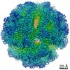 11233MC 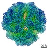 4629C 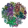 6qsuC M: map data used to model this data C: citing same article ( |
|---|---|
| Similar structure data |


-Links
 PDBj
PDBj

- Assembly
| Deposited unit | 
|
|---|---|
| 1 |
|
-Components
| #1: Protein | Mass: 26645.703 Da / Num. of mol.: 12 Source method: isolated from a genetically manipulated source Source: (gene. exp.) Helicobacter pylori (bacteria) / Gene: ureA, BGL58_00110, D2C78_00705, DD749_00755 / Production host: References: UniProt: A0A293SGE9, UniProt: P14916*PLUS, urease #2: Protein | Mass: 61832.531 Da / Num. of mol.: 12 Source method: isolated from a genetically manipulated source Source: (gene. exp.) Helicobacter pylori (bacteria)Gene: ureC, ureB, ACM26_03100, ACM31_01665, AEY53_07315, AEY54_04500, BB390_03210, BB400_00890, BB458_02040, BB472_00115, BB483_06885, BCM300_00080, BGL59_06905, BGL62_00955, BGL74_08355, BGL78_ ...Gene: ureC, ureB, ACM26_03100, ACM31_01665, AEY53_07315, AEY54_04500, BB390_03210, BB400_00890, BB458_02040, BB472_00115, BB483_06885, BCM300_00080, BGL59_06905, BGL62_00955, BGL74_08355, BGL78_03465, BGL80_00980, BZK19_03120, BZK22_02015, BZK28_01040, C2R48_07340, C2R49_05480, C2R51_07515, C2R54_00600, C2R57_07640, C2R76_03750, C2R81_00275, C2R85_01915, C2R92_02560, CV727_07335, CV730_01155, D2C74_07740, D2C77_03985, D2C79_06860, DD741_00890, DD749_00750, DD779_00665, DD783_00075, DDP32_00750, DDP35_00500, EC528_04020, EC532_02160, EC572_01665, EC592_02340, EC594_04345, EC598_01685, ECB88_00905, ECB90_01230, ECC11_01355, ECC14_06065, ECC20_02355, ECC23_04175, ECC26_01225, ECC27_04455, ECC29_00335, ECC38_01340, ECC45_00115, ECE48_05900, EDB75_03310, EPC84_05055, HPY1198_04550, NCTC13207_01207, NCTC13338_01433, NCTC13345_00234 Production host: References: UniProt: A0A086RWB6, UniProt: P69996*PLUS, urease #3: Chemical | ChemComp-NI /   Mass: 58.693 Da / Num. of mol.: 24 / Source method: obtained synthetically / Formula: Ni Mass: 58.693 Da / Num. of mol.: 24 / Source method: obtained synthetically / Formula: Ni#4: Chemical | ChemComp-DJM /   Mass: 277.342 Da / Num. of mol.: 12 / Source method: obtained synthetically / Formula: C13H15N3O2S Mass: 277.342 Da / Num. of mol.: 12 / Source method: obtained synthetically / Formula: C13H15N3O2S#5: Water | ChemComp-HOH / |  Mass: 18.015 Da / Num. of mol.: 3191 / Source method: isolated from a natural source / Formula: H2O Mass: 18.015 Da / Num. of mol.: 3191 / Source method: isolated from a natural source / Formula: H2OHas ligand of interest | Y | Has protein modification | Y | |
|---|
-Experimental details
-Experiment
| Experiment | Method: ELECTRON MICROSCOPY |
|---|---|
| EM experiment | Aggregation state: PARTICLE / 3D reconstruction method: single particle reconstruction |
- Sample preparation
| Component | Name: 1.1 MDa Helicobacter pylori Urease / Type: COMPLEX / Entity ID: #1-#2 / Source: RECOMBINANT | ||||||||||||||||
|---|---|---|---|---|---|---|---|---|---|---|---|---|---|---|---|---|---|
| Molecular weight | Value: 1.1 MDa / Experimental value: NO | ||||||||||||||||
| Source (natural) | Organism: Helicobacter pylori (bacteria) / Strain: UMAB41 / Cellular location: Cytoplasm | ||||||||||||||||
| Source (recombinant) | Organism: | ||||||||||||||||
| Buffer solution | pH: 8 | ||||||||||||||||
| Buffer component |
| ||||||||||||||||
| Specimen | Conc.: 2 mg/ml / Embedding applied: NO / Shadowing applied: NO / Staining applied: NO / Vitrification applied: YES | ||||||||||||||||
| Vitrification | Instrument: FEI VITROBOT MARK IV / Cryogen name: ETHANE / Humidity: 70 % / Chamber temperature: 283 K |
- Electron microscopy imaging
| Experimental equipment |  Model: Titan Krios / Image courtesy: FEI Company |
|---|---|
| Microscopy | Model: FEI TITAN KRIOS |
| Electron gun | Electron source:  FIELD EMISSION GUN / Accelerating voltage: 300 kV / Illumination mode: FLOOD BEAM FIELD EMISSION GUN / Accelerating voltage: 300 kV / Illumination mode: FLOOD BEAM |
| Electron lens | Mode: BRIGHT FIELD |
| Image recording | Electron dose: 50 e/Å2 / Film or detector model: GATAN K2 SUMMIT (4k x 4k) / Num. of grids imaged: 1 / Num. of real images: 956 / Details: Used 718 movies |
- Processing
| Software | Name: PHENIX / Version: 1.18.1_3865: / Classification: refinement | ||||||||||||||||||||||||||||||||
|---|---|---|---|---|---|---|---|---|---|---|---|---|---|---|---|---|---|---|---|---|---|---|---|---|---|---|---|---|---|---|---|---|---|
| EM software |
| ||||||||||||||||||||||||||||||||
| CTF correction | Details: Relion 3.0 beta per image and per particle / Type: NONE | ||||||||||||||||||||||||||||||||
| Particle selection | Num. of particles selected: 203000 | ||||||||||||||||||||||||||||||||
| Symmetry | Point symmetry: T (tetrahedral) | ||||||||||||||||||||||||||||||||
| 3D reconstruction | Resolution: 2 Å / Resolution method: FSC 0.5 CUT-OFF / Num. of particles: 187461 / Details: Relion was used for resconstruction / Num. of class averages: 1 / Symmetry type: POINT | ||||||||||||||||||||||||||||||||
| Atomic model building | Protocol: RIGID BODY FIT / Space: REAL | ||||||||||||||||||||||||||||||||
| Atomic model building | 3D fitting-ID: 1 / Accession code: 1E9Z / Initial refinement model-ID: 1 / PDB-ID: 1E9Z / Source name: PDB / Type: experimental model
| ||||||||||||||||||||||||||||||||
| Refine LS restraints |
|