 Movie
Movie Controller


+ Open data

- Basic information
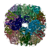
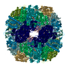
| Entry | Database: PDB / ID: 1e9z | ||||||
|---|---|---|---|---|---|---|---|
| Title | Crystal structure of Helicobacter pylori urease | ||||||
 Components Components |
| ||||||
 Keywords Keywords | HYDROLASE | ||||||
| Function / homology |  Function and homology information Function and homology informationurease complex / urease / urease activity / urea catabolic process / nickel cation binding / cytoplasm Similarity search - Function | ||||||
| Biological species |   HELICOBACTER PYLORI (bacteria) HELICOBACTER PYLORI (bacteria) | ||||||
| Method |  X-RAY DIFFRACTION / MOLECULAR REPLACEMENT / Resolution: 3 Å X-RAY DIFFRACTION / MOLECULAR REPLACEMENT / Resolution: 3 Å | ||||||
 Authors Authors | Ha, N.-C. / Oh, S.-T. / Oh, B.-H. | ||||||
 Citation Citation | Journal: Nat.Struct.Biol. / Year: 2001 Title: Supramolecular Assembly and Acid Resistance of Helicobacter Pylori Urease Authors: Ha, N.-C. / Oh, S.-T. / Sung, J.Y. / Cha, K.-A. / Hyung Lee, M. / Oh, B.-H. | ||||||
| History |
|
- Structure visualization
| Structure viewer | Molecule: MolmilJmol/JSmol |
|---|
- Downloads & links
-Download
| PDBx/mmCIF format | 1e9z.cif.gz | 166.8 KB | Display | PDBx/mmCIF format |
|---|---|---|---|---|
| PDB format | pdb1e9z.ent.gz | 130.1 KB | Display | PDB format |
| PDBx/mmJSON format | 1e9z.json.gz | Tree view | PDBx/mmJSON format | |
| Others |  Other downloads Other downloads |
-Validation report
| Arichive directory | https://data.pdbj.org/pub/pdb/validation_reports/e9/1e9zftp://data.pdbj.org/pub/pdb/validation_reports/e9/1e9z | HTTPS FTP |
|---|
-Related structure data
| Related structure data | 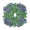 1e9yC  1kau C: citing same article ( S: Starting model for refinement |
|---|---|
| Similar structure data |


-Links
 PDBj
PDBj

- Assembly
| Deposited unit | 
| ||||||||
|---|---|---|---|---|---|---|---|---|---|
| 1 | x 12
| ||||||||
| Unit cell |
|
-Components
| #1: Protein | Mass: 26571.662 Da / Num. of mol.: 1 Source method: isolated from a genetically manipulated source Source: (gene. exp.) HELICOBACTER PYLORI (bacteria) / Production host: | ||
|---|---|---|---|
| #2: Protein | Mass: 61745.422 Da / Num. of mol.: 1 Source method: isolated from a genetically manipulated source Source: (gene. exp.) HELICOBACTER PYLORI (bacteria) / Production host: | ||
| #3: Chemical |   Mass: 58.693 Da / Num. of mol.: 2 / Source method: obtained synthetically / Formula: Ni Mass: 58.693 Da / Num. of mol.: 2 / Source method: obtained synthetically / Formula: Ni#4: Water | ChemComp-HOH / |  Mass: 18.015 Da / Num. of mol.: 78 / Source method: isolated from a natural source / Formula: H2O Mass: 18.015 Da / Num. of mol.: 78 / Source method: isolated from a natural source / Formula: H2O |
-Experimental details
-Experiment
| Experiment | Method: X-RAY DIFFRACTION / Number of used crystals: 1 |
|---|
- Sample preparation
| Crystal | Density Matthews: 2.66 Å3/Da / Density % sol: 51 % | ||||||||||||||||||||
|---|---|---|---|---|---|---|---|---|---|---|---|---|---|---|---|---|---|---|---|---|---|
| Crystal grow | pH: 6.5 / Details: pH 6.50 | ||||||||||||||||||||
| Crystal grow | *PLUS Temperature: 12 ℃ / Method: vapor diffusion, hanging drop / Details: used macroseeding | ||||||||||||||||||||
| Components of the solutions | *PLUS
|
-Data collection
| Diffraction | Mean temperature: 100 K |
|---|---|
| Diffraction source | Source: ROTATING ANODE / Type: MACSCIENCE M18X / Wavelength: 1.5418 |
| Detector | Type: MACSCIENCE / Detector: IMAGE PLATE |
| Radiation | Monochromator: GRAPHITE / Protocol: SINGLE WAVELENGTH / Monochromatic (M) / Laue (L): M / Scattering type: x-ray |
| Radiation wavelength | Wavelength: 1.5418 Å / Relative weight: 1 |
| Reflection | Resolution: 3→20 Å / Num. obs: 21702 / % possible obs: 95.3 % / Observed criterion σ(I): 1 / Redundancy: 20 % / Rmerge(I) obs: 0.115 / Rsym value: 0.115 / Net I/σ(I): 5.7 |
| Reflection | *PLUS Highest resolution: 2.8 Å / Lowest resolution: 20 Å / Num. obs: 23292 / % possible obs: 89.1 % / Num. measured all: 393095 / Rmerge(I) obs: 0.114 |
| Reflection shell | *PLUS % possible obs: 75.8 % / Rmerge(I) obs: 0.317 |
- Processing
| Software |
| ||||||||||||||||||||||||||||||||||||||||||||||||||||||||||||
|---|---|---|---|---|---|---|---|---|---|---|---|---|---|---|---|---|---|---|---|---|---|---|---|---|---|---|---|---|---|---|---|---|---|---|---|---|---|---|---|---|---|---|---|---|---|---|---|---|---|---|---|---|---|---|---|---|---|---|---|---|---|
| Refinement | Method to determine structure: MOLECULAR REPLACEMENT Starting model: PDB ENTRY 1KAU 1kau Resolution: 3→20 Å / Cross valid method: THROUGHOUT / σ(F): 1
| ||||||||||||||||||||||||||||||||||||||||||||||||||||||||||||
| Refinement step | Cycle: LAST / Resolution: 3→20 Å
| ||||||||||||||||||||||||||||||||||||||||||||||||||||||||||||
| Refine LS restraints |
| ||||||||||||||||||||||||||||||||||||||||||||||||||||||||||||
| Software | *PLUS Name: CNS / Classification: refinement | ||||||||||||||||||||||||||||||||||||||||||||||||||||||||||||
| Refinement | *PLUS Highest resolution: 2.8 Å / Lowest resolution: 40 Å / σ(F): 1 / Rfactor obs: 0.206 / Rfactor Rfree: 0.287 | ||||||||||||||||||||||||||||||||||||||||||||||||||||||||||||
| Solvent computation | *PLUS | ||||||||||||||||||||||||||||||||||||||||||||||||||||||||||||
| Displacement parameters | *PLUS |
