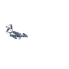
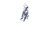
Movie
Movie Controller


+ Open data

- Basic information
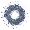
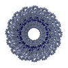
| Entry | Database: PDB / ID: 6r7m | |||||||||
|---|---|---|---|---|---|---|---|---|---|---|
| Title | Tobacco Mosaic Virus (TMV) | |||||||||
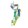
 Components Components | Capsid protein | |||||||||
 Keywords Keywords | VIRUS / tobacco / mosaic / TMV / cryo-EM / cryoWriter / microfluidic | |||||||||
| Function / homology |  Function and homology information Function and homology informationhelical viral capsid / structural molecule activity / identical protein binding Similarity search - Function | |||||||||
| Biological species |  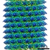 Tobacco mosaic virus Tobacco mosaic virus | |||||||||
| Method | ELECTRON MICROSCOPY / helical reconstruction / cryo EM / Resolution: 1.92 Å | |||||||||
 Authors Authors | Schmidli, C. / Albiez, S. / Rima, L. / Righetto, R. / Mohammed, I. / Oliva, P. / Kovacik, L. / Stahlberg, H. / Braun, T. | |||||||||
| Funding support |  Switzerland, 2items Switzerland, 2items
| |||||||||
 Citation Citation | Journal: Proc Natl Acad Sci U S A / Year: 2019 Title: Microfluidic protein isolation and sample preparation for high-resolution cryo-EM. Authors: Claudio Schmidli / Stefan Albiez / Luca Rima / Ricardo Righetto / Inayatulla Mohammed / Paolo Oliva / Lubomir Kovacik / Henning Stahlberg / Thomas Braun / Abstract: High-resolution structural information is essential to understand protein function. Protein-structure determination needs a considerable amount of protein, which can be challenging to produce, often ...High-resolution structural information is essential to understand protein function. Protein-structure determination needs a considerable amount of protein, which can be challenging to produce, often involving harsh and lengthy procedures. In contrast, the several thousand to a few million protein particles required for structure determination by cryogenic electron microscopy (cryo-EM) can be provided by miniaturized systems. Here, we present a microfluidic method for the rapid isolation of a target protein and its direct preparation for cryo-EM. Less than 1 μL of cell lysate is required as starting material to solve the atomic structure of the untagged, endogenous human 20S proteasome. Our work paves the way for high-throughput structure determination of proteins from minimal amounts of cell lysate and opens more opportunities for the isolation of sensitive, endogenous protein complexes. | |||||||||
| History |
|
- Structure visualization
| Movie |
Movie viewer |
|---|---|
| Structure viewer | Molecule: MolmilJmol/JSmol |
- Downloads & links
-Download
| PDBx/mmCIF format | 6r7m.cif.gz | 38.9 KB | Display | PDBx/mmCIF format |
|---|---|---|---|---|
| PDB format | pdb6r7m.ent.gz | 25.9 KB | Display | PDB format |
| PDBx/mmJSON format | 6r7m.json.gz | Tree view | PDBx/mmJSON format | |
| Others |  Other downloads Other downloads |
-Validation report
| Arichive directory | https://data.pdbj.org/pub/pdb/validation_reports/r7/6r7mftp://data.pdbj.org/pub/pdb/validation_reports/r7/6r7m | HTTPS FTP |
|---|
-Related structure data
| Related structure data | 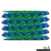 4628MC  4738C  6r70C M: map data used to model this data C: citing same article ( |
|---|---|
| Similar structure data |
-Links
 PDBj
PDBj
- Assembly
| Deposited unit | 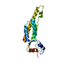
|
|---|---|
| 1 | x 40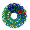
|
| 2 |
|
| Symmetry | Helical symmetry: (Circular symmetry: 1 / Dyad axis: no / N subunits divisor: 1 / Num. of operations: 40 / Rise per n subunits: 1.41 Å / Rotation per n subunits: 22.03 °) |
-Components
| #1: Protein | Mass: 17091.998 Da / Num. of mol.: 1 / Source method: isolated from a natural source / Source: (natural) Tobacco mosaic virus / References: UniProt: A0A348G6U1, UniProt: P69687*PLUS |
|---|
-Experimental details
-Experiment
| Experiment | Method: ELECTRON MICROSCOPY |
|---|---|
| EM experiment | Aggregation state: HELICAL ARRAY / 3D reconstruction method: helical reconstruction |
- Sample preparation
| Component | Name: Tobacco mosaic virus / Type: VIRUS / Entity ID: all / Source: NATURAL |
|---|---|
| Molecular weight | Value: 39.5 MDa / Experimental value: NO |
| Source (natural) | Organism: Tobacco mosaic virus |
| Details of virus | Empty: NO / Enveloped: NO / Isolate: STRAIN / Type: VIRUS-LIKE PARTICLE |
| Natural host | Organism: Nicotiana tabacum |
| Virus shell | Name: CP / Diameter: 180 nm |
| Buffer solution | pH: 7.4 |
| Specimen | Conc.: 1 mg/ml / Embedding applied: NO / Shadowing applied: NO / Staining applied: NO / Vitrification applied: YES |
| Specimen support | Grid material: COPPER / Grid mesh size: 400 divisions/in. / Grid type: Quantifoil R1.2/1.3 |
| Vitrification | Cryogen name: ETHANE / Details: CryoWriter |
- Electron microscopy imaging
| Experimental equipment |  Model: Titan Krios / Image courtesy: FEI Company |
|---|---|
| Microscopy | Model: FEI TITAN KRIOS |
| Electron gun | Electron source:  FIELD EMISSION GUN / Accelerating voltage: 300 kV / Illumination mode: FLOOD BEAM FIELD EMISSION GUN / Accelerating voltage: 300 kV / Illumination mode: FLOOD BEAM |
| Electron lens | Mode: BRIGHT FIELD / Cs: 2.7 mm / C2 aperture diameter: 70 µm / Alignment procedure: COMA FREE |
| Specimen holder | Cryogen: NITROGEN / Specimen holder model: FEI TITAN KRIOS AUTOGRID HOLDER |
| Image recording | Average exposure time: 6 sec. / Electron dose: 72 e/Å2 / Detector mode: SUPER-RESOLUTION / Film or detector model: GATAN K2 QUANTUM (4k x 4k) / Num. of grids imaged: 1 / Num. of real images: 523 |
| Image scans | Width: 7676 / Height: 7420 / Movie frames/image: 30 / Used frames/image: 0-30 |
- Processing
| EM software |
| |||||||||||||||||||||||||||||||||||
|---|---|---|---|---|---|---|---|---|---|---|---|---|---|---|---|---|---|---|---|---|---|---|---|---|---|---|---|---|---|---|---|---|---|---|---|---|
| CTF correction | Type: PHASE FLIPPING AND AMPLITUDE CORRECTION | |||||||||||||||||||||||||||||||||||
| Helical symmerty | Angular rotation/subunit: 22.03 ° / Axial rise/subunit: 1.41 Å / Axial symmetry: C1 | |||||||||||||||||||||||||||||||||||
| Particle selection | Num. of particles selected: 2676 | |||||||||||||||||||||||||||||||||||
| 3D reconstruction | Resolution: 1.92 Å / Resolution method: FSC 0.143 CUT-OFF / Num. of particles: 52776 / Algorithm: BACK PROJECTION / Symmetry type: HELICAL | |||||||||||||||||||||||||||||||||||
| Atomic model building | PDB-ID: 6I5A Accession code: 6I5A / Source name: PDB / Type: experimental model |