 Movie
Movie Controller


+ Open data

- Basic information
| Entry | Database: PDB / ID: 6i5a | ||||||
|---|---|---|---|---|---|---|---|
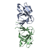
| Title | Tobacco Mosaic Virus | ||||||
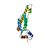
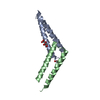
 Components Components | Capsid protein | ||||||
 Keywords Keywords | VIRUS / TMV / helical virus / coat protein / plant pathogen | ||||||
| Function / homology |  Function and homology information Function and homology informationhelical viral capsid / structural molecule activity / identical protein binding Similarity search - Function | ||||||
| Biological species |  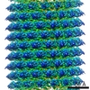 Tobacco mosaic virus Tobacco mosaic virus | ||||||
| Method | ELECTRON MICROSCOPY / helical reconstruction / cryo EM / Resolution: 2.3 Å | ||||||
 Authors Authors | Song, B. / Flegler, V. / Makbul, C. / Rasmussen, T. / Bottcher, B. | ||||||
| Funding support |  Germany, 1items Germany, 1items
| ||||||
 Citation Citation | Journal: Ultramicroscopy / Year: 2019 Title: Capabilities of the Falcon III detector for single-particle structure determination. Authors: Boyuan Song / Julian Lenhart / Vanessa Judith Flegler / Cihan Makbul / Tim Rasmussen / Bettina Böttcher / Abstract: Direct electron detectors are an essential asset for the resolution revolution in electron cryo microscopy of biological objects. The direct detectors provide two modes of data acquisition; the ...Direct electron detectors are an essential asset for the resolution revolution in electron cryo microscopy of biological objects. The direct detectors provide two modes of data acquisition; the counting mode in which single electrons are counted, and the integrating mode in which the signal that arises from the incident electrons is integrated. While counting mode leads to far higher detective quantum efficiency at all spatial frequencies, the integrating mode enables faster data acquisition at higher exposure rates. For optimal throughput at best possible resolution it is important to understand when the better performance in counting mode becomes essential for solving a structure and when the lower detective quantum efficiency in integrating mode can be compensated by increasing the number of particles in the data set. Here, we provide a case study of the Falcon III camera, which has counting mode capability at exposure rates of <0.9 e/Px² and integrating mode capability at exposure rates above 10 e/Px². We found that counting mode gives better resolution for medium sized complexes such as the β-galactosidase (465 kDa) (2.2 Å, 97% of Nyquist vs. 2.4 Å, 89% of Nyquist) with data sets of similar size. However, for larger particles such as Hepatitis B virus capsid like particles (4.8 MDa) we did not find any resolution gain in counting mode. | ||||||
| History |
|
- Structure visualization
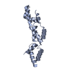
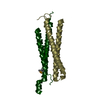
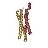
| Movie |
Movie viewer |
|---|---|
| Structure viewer | Molecule: MolmilJmol/JSmol |
- Downloads & links
-Download
| PDBx/mmCIF format | 6i5a.cif.gz | 41.4 KB | Display | PDBx/mmCIF format |
|---|---|---|---|---|
| PDB format | pdb6i5a.ent.gz | 27.9 KB | Display | PDB format |
| PDBx/mmJSON format | 6i5a.json.gz | Tree view | PDBx/mmJSON format | |
| Others |  Other downloads Other downloads |
-Validation report
| Arichive directory | https://data.pdbj.org/pub/pdb/validation_reports/i5/6i5aftp://data.pdbj.org/pub/pdb/validation_reports/i5/6i5a | HTTPS FTP |
|---|
-Related structure data
| Related structure data | 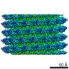 4413MC 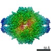 4414C 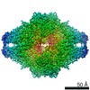 4415C 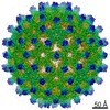 4416C 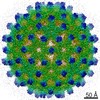 4417C M: map data used to model this data C: citing same article ( |
|---|---|
| Similar structure data |


-Links
 PDBj
PDBj
- Assembly
| Deposited unit | 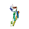
|
|---|---|
| 1 |
|
-Components
| #1: Protein | Mass: 17636.621 Da / Num. of mol.: 1 / Source method: isolated from a natural source / Source: (natural) Tobacco mosaic virus (strain vulgare) / Strain: vulgare / References: UniProt: P69687 |
|---|
-Experimental details
-Experiment
| Experiment | Method: ELECTRON MICROSCOPY |
|---|---|
| EM experiment | Aggregation state: HELICAL ARRAY / 3D reconstruction method: helical reconstruction |
- Sample preparation
| Component | Name: Tobacco mosaic virus / Type: VIRUS / Entity ID: all / Source: NATURAL |
|---|---|
| Molecular weight | Value: 40 MDa / Experimental value: NO |
| Source (natural) | Organism: Tobacco mosaic virus |
| Details of virus | Empty: NO / Enveloped: NO / Isolate: STRAIN / Type: VIRION |
| Natural host | Organism: Nicotiana sp. |
| Virus shell | Diameter: 180 nm |
| Buffer solution | pH: 7 |
| Specimen | Conc.: 10 mg/ml / Embedding applied: NO / Shadowing applied: NO / Staining applied: NO / Vitrification applied: YES |
| Specimen support | Grid material: COPPER / Grid mesh size: 400 divisions/in. / Grid type: Quantifoil R1.2/1.3 |
| Vitrification | Instrument: FEI VITROBOT MARK IV / Cryogen name: ETHANE / Humidity: 100 % / Chamber temperature: 277 K Details: Quantifoil 400 mesh copper grids R 1.2/1.3 were used (Quantifoil Micro Tools GmbH, Jena, Germany). Grids were glow discharged in air at a pressure of 2.2x10⁻² Torr for 2 min at medium power ...Details: Quantifoil 400 mesh copper grids R 1.2/1.3 were used (Quantifoil Micro Tools GmbH, Jena, Germany). Grids were glow discharged in air at a pressure of 2.2x10⁻² Torr for 2 min at medium power with a Harrick Plasma cleaner (PDC-002). Subsequently, 3-4 ul of the sample was pipetted onto the glow discharged grids and plunge frozen in liquid Ethane with a Vitrobot IV (FEI). The sample chamber of the Vitrobot was maintained at 4C and 100% humidity. Wait time between sample application and blotting was 45 s, the drain time 0 s, the blot time 3 s and the blot force 0. |
- Electron microscopy imaging
| Experimental equipment |  Model: Titan Krios / Image courtesy: FEI Company |
|---|---|
| Microscopy | Model: FEI TITAN KRIOS |
| Electron gun | Electron source:  FIELD EMISSION GUN / Accelerating voltage: 300 kV / Illumination mode: FLOOD BEAM FIELD EMISSION GUN / Accelerating voltage: 300 kV / Illumination mode: FLOOD BEAM |
| Electron lens | Mode: BRIGHT FIELD / Nominal magnification: 75000 X / Nominal defocus max: 1800 nm / Nominal defocus min: 1000 nm / Calibrated defocus min: 580 nm / Calibrated defocus max: 2120 nm / Cs: 2.7 mm / C2 aperture diameter: 70 µm / Alignment procedure: COMA FREE |
| Specimen holder | Cryogen: NITROGEN / Specimen holder model: FEI TITAN KRIOS AUTOGRID HOLDER / Temperature (max): 80 K / Temperature (min): 79 K |
| Image recording | Average exposure time: 5 sec. / Electron dose: 55 e/Å2 / Detector mode: INTEGRATING / Film or detector model: FEI FALCON III (4k x 4k) / Num. of grids imaged: 1 / Num. of real images: 1001 |
| Image scans | Width: 4096 / Height: 4096 |
- Processing
| EM software |
| ||||||||||||||||||||||||||||||||||||||||||||||||||
|---|---|---|---|---|---|---|---|---|---|---|---|---|---|---|---|---|---|---|---|---|---|---|---|---|---|---|---|---|---|---|---|---|---|---|---|---|---|---|---|---|---|---|---|---|---|---|---|---|---|---|---|
| Image processing | Details: Movies were motion corrected and exposure weighted either with motioncor2 (see tables 1-3 for details). The defocus was determined for the motion corrected and exposure weighted averages with ctffind4 | ||||||||||||||||||||||||||||||||||||||||||||||||||
| CTF correction | Type: PHASE FLIPPING AND AMPLITUDE CORRECTION | ||||||||||||||||||||||||||||||||||||||||||||||||||
| Helical symmerty | Angular rotation/subunit: 22.037 ° / Axial rise/subunit: 1.39615 Å / Axial symmetry: C1 | ||||||||||||||||||||||||||||||||||||||||||||||||||
| Particle selection | Num. of particles selected: 393692 Details: For TMV, some helices were picked manually with the helix picker implemented in relion 2.1. An initial 3D model of these segments was determined using the relion helix option with a cylinder ...Details: For TMV, some helices were picked manually with the helix picker implemented in relion 2.1. An initial 3D model of these segments was determined using the relion helix option with a cylinder as the starting reference. The aligned helical segments after 3D refinement were subjected to 2D classification without alignment. Three of the 2D class averages were selected as templates for automatic picking of the TMV segments in relion. | ||||||||||||||||||||||||||||||||||||||||||||||||||
| 3D reconstruction | Resolution: 2.3 Å / Resolution method: FSC 0.143 CUT-OFF / Num. of particles: 91927 / Algorithm: FOURIER SPACE / Num. of class averages: 1 / Symmetry type: HELICAL | ||||||||||||||||||||||||||||||||||||||||||||||||||
| Atomic model building | B value: 105 / Protocol: AB INITIO MODEL / Space: RECIPROCAL |
