 Movie
Movie Controller

[English] 日本語
 Yorodumi
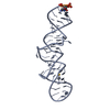
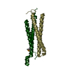
- PDB-5uae: Crystal structure of the coiled-coil domain from Listeria Innocua... -
+ Open data

- Basic information
| Entry | Database: PDB / ID: 5uae | ||||||
|---|---|---|---|---|---|---|---|
| Title | Crystal structure of the coiled-coil domain from Listeria Innocua Phage Integrase (Trigonal Form) | ||||||
 Components Components | Putative integrase | ||||||
 Keywords Keywords | RECOMBINATION / site-specific recombination / coiled-coil | ||||||
| Function / homology |  Function and homology information Function and homology informationDNA strand exchange activity / DNA integration / DNA binding / metal ion binding Similarity search - Function | ||||||
| Biological species |  Listeria innocua (bacteria) Listeria innocua (bacteria) | ||||||
| Method |  X-RAY DIFFRACTION / SYNCHROTRON / MOLECULAR REPLACEMENT / Resolution: 2.75 Å X-RAY DIFFRACTION / SYNCHROTRON / MOLECULAR REPLACEMENT / Resolution: 2.75 Å | ||||||
 Authors Authors | Gupta, K. / Sharp, R. / Yuan, J.B. / Van Duyne, G.D. | ||||||
| Funding support |  United States, 1items United States, 1items
| ||||||
 Citation Citation | Journal: Nucleic Acids Res. / Year: 2017 Title: Coiled-coil interactions mediate serine integrase directionality. Authors: Gupta, K. / Sharp, R. / Yuan, J.B. / Li, H. / Van Duyne, G.D. #1: Journal: Nucleic Acids Res. / Year: 2013Title: Attachment site recognition and regulation of directionality by the serine integrases. Authors: Rutherford, K. / Yuan, P. / Perry, K. / Sharp, R. / Van Duyne, G.D. | ||||||
| History |
|
- Structure visualization
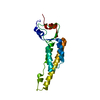
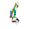
| Structure viewer | Molecule: MolmilJmol/JSmol |
|---|
- Downloads & links
-Download
| PDBx/mmCIF format | 5uae.cif.gz | 69.3 KB | Display | PDBx/mmCIF format |
|---|---|---|---|---|
| PDB format | pdb5uae.ent.gz | 52.4 KB | Display | PDB format |
| PDBx/mmJSON format | 5uae.json.gz | Tree view | PDBx/mmJSON format | |
| Others |  Other downloads Other downloads |
-Validation report
| Arichive directory | https://data.pdbj.org/pub/pdb/validation_reports/ua/5uaeftp://data.pdbj.org/pub/pdb/validation_reports/ua/5uae | HTTPS FTP |
|---|
-Related structure data
| Related structure data |  5u96C 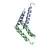 5udoC  4kisS C: citing same article ( S: Starting model for refinement |
|---|---|
| Similar structure data |


-Links
 PDBj
PDBj


- Assembly
| Deposited unit | 
| ||||||||
|---|---|---|---|---|---|---|---|---|---|
| 1 | 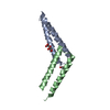
| ||||||||
| 2 | 
| ||||||||
| Unit cell |
|
-Components
| #1: Protein | Mass: 7364.188 Da / Num. of mol.: 4 / Fragment: Coiled Coil Domain (UNP residues 344-405) Source method: isolated from a genetically manipulated source Source: (gene. exp.) Listeria innocua (bacteria) / Gene: int / Production host: #2: Chemical | ChemComp-FLC /   Mass: 189.100 Da / Num. of mol.: 5 / Source method: obtained synthetically / Formula: C6H5O7 Mass: 189.100 Da / Num. of mol.: 5 / Source method: obtained synthetically / Formula: C6H5O7#3: Water | ChemComp-HOH / |  Mass: 18.015 Da / Num. of mol.: 249 / Source method: isolated from a natural source / Formula: H2O Mass: 18.015 Da / Num. of mol.: 249 / Source method: isolated from a natural source / Formula: H2O |
|---|
-Experimental details
-Experiment
| Experiment | Method: X-RAY DIFFRACTION / Number of used crystals: 1 |
|---|
- Sample preparation
| Crystal | Density Matthews: 5.75 Å3/Da / Density % sol: 78.6 % |
|---|---|
| Crystal grow | Temperature: 294.15 K / Method: vapor diffusion, hanging drop / pH: 7.4 / Details: 0.8-1.2 M Na Citrate |
-Data collection
| Diffraction | Mean temperature: 100 K |
|---|---|
| Diffraction source | Source: SYNCHROTRON / Site: CHESS / Beamline: F1 / Wavelength: 1 Å |
| Detector | Type: DECTRIS PILATUS 6M / Detector: PIXEL / Date: May 26, 2016 |
| Radiation | Protocol: SINGLE WAVELENGTH / Monochromatic (M) / Laue (L): M / Scattering type: x-ray |
| Radiation wavelength | Wavelength: 1 Å / Relative weight: 1 |
| Reflection | Resolution: 2.75→37.716 Å / Num. obs: 69598 / % possible obs: 99.7 % / Redundancy: 4.1 % / Biso Wilson estimate: 91.1 Å2 / Rmerge(I) obs: 0.052 / Net I/σ(I): 15.3 |
| Reflection shell | Resolution: 2.75→2.848 Å / Redundancy: 3.7 % / Rmerge(I) obs: 0.623 / Mean I/σ(I) obs: 1.8 / % possible all: 99 |
- Processing
| Software |
| |||||||||||||||||||||||||||||||||||||||||||||||||||||||||||||||||||||||||||||||||||||||||||
|---|---|---|---|---|---|---|---|---|---|---|---|---|---|---|---|---|---|---|---|---|---|---|---|---|---|---|---|---|---|---|---|---|---|---|---|---|---|---|---|---|---|---|---|---|---|---|---|---|---|---|---|---|---|---|---|---|---|---|---|---|---|---|---|---|---|---|---|---|---|---|---|---|---|---|---|---|---|---|---|---|---|---|---|---|---|---|---|---|---|---|---|---|
| Refinement | Method to determine structure: MOLECULAR REPLACEMENT Starting model: 4KIS Resolution: 2.75→37.716 Å / Cross valid method: FREE R-VALUE / σ(F): 2 / Phase error: 28.78 / Stereochemistry target values: TWIN_LSQ_F
| |||||||||||||||||||||||||||||||||||||||||||||||||||||||||||||||||||||||||||||||||||||||||||
| Solvent computation | Shrinkage radii: 0.9 Å / VDW probe radii: 1.11 Å / Solvent model: FLAT BULK SOLVENT MODEL | |||||||||||||||||||||||||||||||||||||||||||||||||||||||||||||||||||||||||||||||||||||||||||
| Refinement step | Cycle: LAST / Resolution: 2.75→37.716 Å
| |||||||||||||||||||||||||||||||||||||||||||||||||||||||||||||||||||||||||||||||||||||||||||
| Refine LS restraints |
| |||||||||||||||||||||||||||||||||||||||||||||||||||||||||||||||||||||||||||||||||||||||||||
| LS refinement shell |
|
