 Movie
Movie Controller

[English] 日本語
 Yorodumi
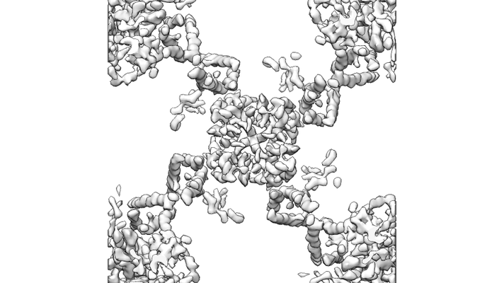
- EMDB-4517: MloK1 map from single particle analysis of 2D crystals, class 6 (... -
+ Open data

- Basic information
| Entry | Database: EMDB / ID: EMD-4517 | |||||||||
|---|---|---|---|---|---|---|---|---|---|---|
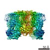
| Title | MloK1 map from single particle analysis of 2D crystals, class 6 (intermediate compact conformation) | |||||||||
 Map data Map data | ||||||||||
 Sample Sample |
| |||||||||
 Keywords Keywords | Voltage-gated potassium channel / Cyclic nucleotide-binding domain / ion channel / ion transport / MEMBRANE PROTEIN | |||||||||
| Function / homology |  Function and homology information Function and homology informationintracellularly cyclic nucleotide-activated monoatomic cation channel activity / potassium channel activity / cAMP binding / protein-containing complex binding / identical protein binding / plasma membrane Similarity search - Function | |||||||||
| Biological species |  Mesorhizobium loti MAFF303099 (bacteria) Mesorhizobium loti MAFF303099 (bacteria) | |||||||||
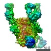
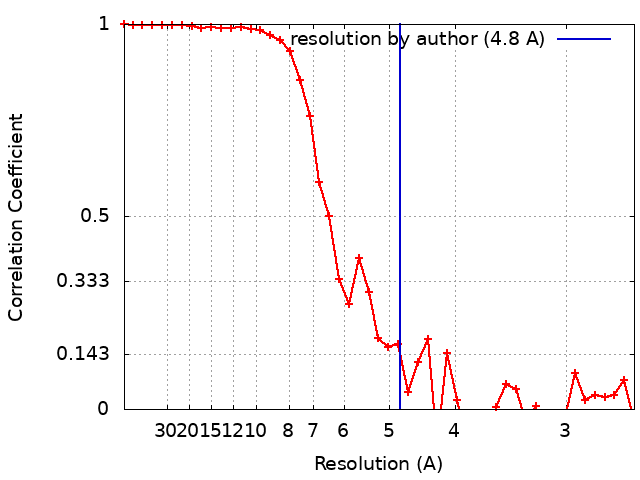
| Method | single particle reconstruction / cryo EM / Resolution: 4.8 Å | |||||||||
 Authors Authors | Righetto R / Biyani N | |||||||||
| Funding support |  Switzerland, 1 items Switzerland, 1 items
| |||||||||
 Citation Citation | Journal: Nat Commun / Year: 2019 Title: Retrieving high-resolution information from disordered 2D crystals by single-particle cryo-EM. Authors: Ricardo D Righetto / Nikhil Biyani / Julia Kowal / Mohamed Chami / Henning Stahlberg / Abstract: Electron crystallography can reveal the structure of membrane proteins within 2D crystals under close-to-native conditions. High-resolution structural information can only be reached if crystals are ...Electron crystallography can reveal the structure of membrane proteins within 2D crystals under close-to-native conditions. High-resolution structural information can only be reached if crystals are perfectly flat and highly ordered. In practice, such crystals are difficult to obtain. Available image unbending algorithms correct for disorder, but only perform well on images of non-tilted, flat crystals, while out-of-plane distortions are not addressed. Here, we present an approach that employs single-particle refinement procedures to locally unbend crystals in 3D. With this method, density maps of the MloK1 potassium channel with a resolution of 4 Å were obtained from images of 2D crystals that do not diffract beyond 10 Å. Furthermore, 3D classification allowed multiple structures to be resolved, revealing a series of MloK1 conformations within a single 2D crystal. This conformational heterogeneity explains the poor diffraction observed and is related to channel function. The approach is implemented in the FOCUS package. | |||||||||
| History |
|
- Structure visualization
| Movie |
Movie viewer |
|---|---|
| Structure viewer | EM map: SurfViewMolmilJmol/JSmol |
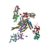
| Supplemental images |
- Downloads & links
-EMDB archive
| Map data | emd_4517.map.gz | 4 MB | EMDB map data format | |
|---|---|---|---|---|
| Header (meta data) | emd-4517-v30.xmlemd-4517.xml | 17.4 KB 17.4 KB | Display Display | EMDB header |
| FSC (resolution estimation) | emd_4517_fsc.xml | 3.8 KB | Display | FSC data file |
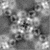
| Images |  emd_4517.png emd_4517.png | 83.9 KB | ||
| Masks | emd_4517_msk_1.map | 4.3 MB | Mask map | |
| Filedesc metadata | emd-4517.cif.gz | 6.5 KB | ||
| Others | emd_4517_half_map_1.map.gzemd_4517_half_map_2.map.gz | 4 MB 4 MB | ||
| Archive directory |  http://ftp.pdbj.org/pub/emdb/structures/EMD-4517ftp://ftp.pdbj.org/pub/emdb/structures/EMD-4517 http://ftp.pdbj.org/pub/emdb/structures/EMD-4517ftp://ftp.pdbj.org/pub/emdb/structures/EMD-4517 | HTTPS FTP |
-Related structure data
| Related structure data | 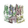 6qd2MC  4432C  4439C 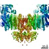 4441C 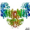 4513C 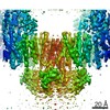 4514C 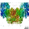 4515C 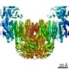 4516C 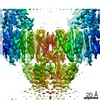 4518C 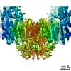 4519C 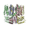 6i9dC 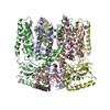 6iaxC 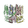 6qcyC 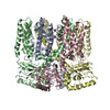 6qczC 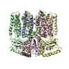 6qd0C 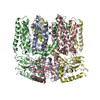 6qd1C 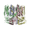 6qd3C 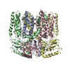 6qd4C M: atomic model generated by this map C: citing same article ( |
|---|---|
| Similar structure data |
-Links
| EMDB pages | EMDB (EBI/PDBe) / EMDataResource |
|---|---|
| Related items in Molecule of the Month |


-Map
| File | Download / File: emd_4517.map.gz / Format: CCP4 / Size: 4.3 MB / Type: IMAGE STORED AS FLOATING POINT NUMBER (4 BYTES) | ||||||||||||||||||||||||||||||||||||||||||||||||||||||||||||
|---|---|---|---|---|---|---|---|---|---|---|---|---|---|---|---|---|---|---|---|---|---|---|---|---|---|---|---|---|---|---|---|---|---|---|---|---|---|---|---|---|---|---|---|---|---|---|---|---|---|---|---|---|---|---|---|---|---|---|---|---|---|
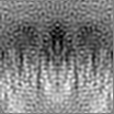
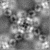
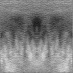
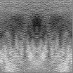
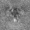
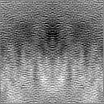
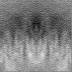
| Projections & slices | Image control
Images are generated by Spider. | ||||||||||||||||||||||||||||||||||||||||||||||||||||||||||||
| Voxel size | X=Y=Z: 1.3 Å | ||||||||||||||||||||||||||||||||||||||||||||||||||||||||||||
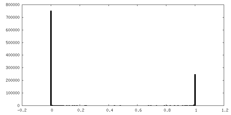
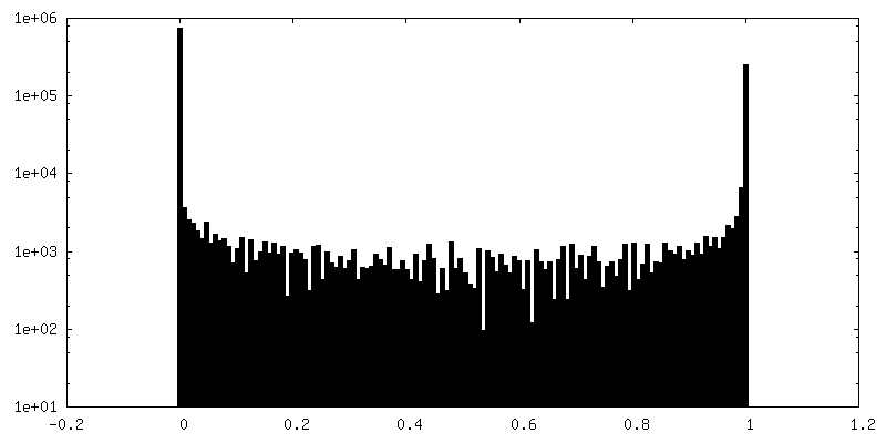
| Density |
| ||||||||||||||||||||||||||||||||||||||||||||||||||||||||||||
| Symmetry | Space group: 1 | ||||||||||||||||||||||||||||||||||||||||||||||||||||||||||||
| Details | EMDB XML:
CCP4 map header:
| ||||||||||||||||||||||||||||||||||||||||||||||||||||||||||||
 Z (Sec.)
 Y (Row.)
 X (Col.)


-Supplemental data
-Mask #1
| File | emd_4517_msk_1.map | ||||||||||||
|---|---|---|---|---|---|---|---|---|---|---|---|---|---|
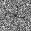
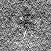
| Projections & Slices |
| ||||||||||||
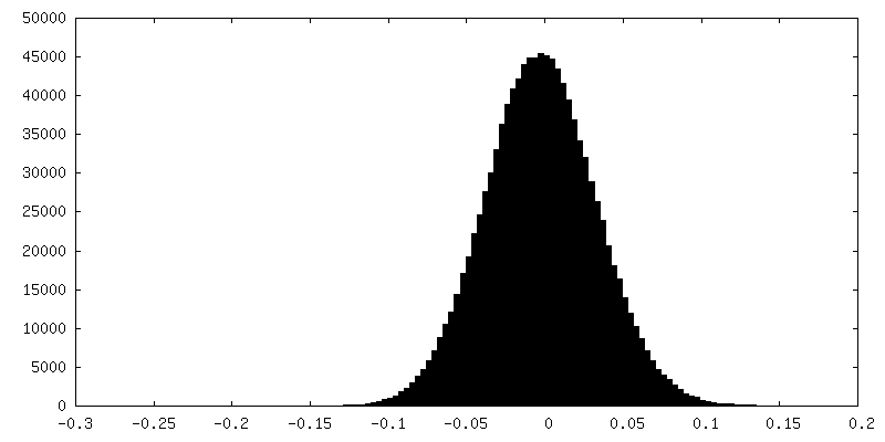
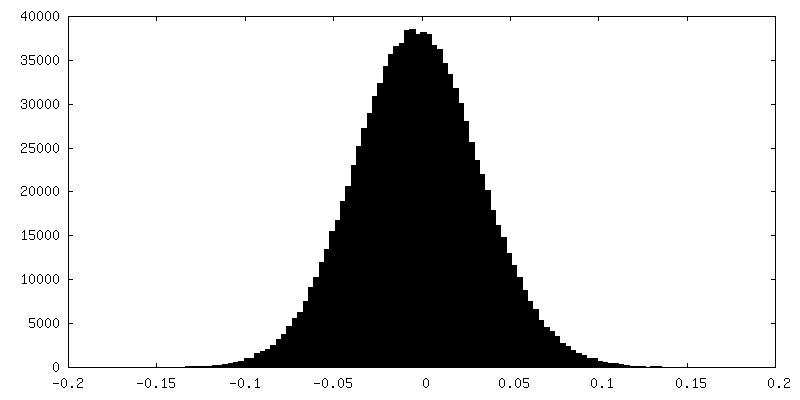
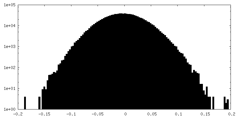
| Density Histograms |


-Half map: #2
| File | emd_4517_half_map_1.map | ||||||||||||
|---|---|---|---|---|---|---|---|---|---|---|---|---|---|
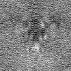
| Projections & Slices |
| ||||||||||||
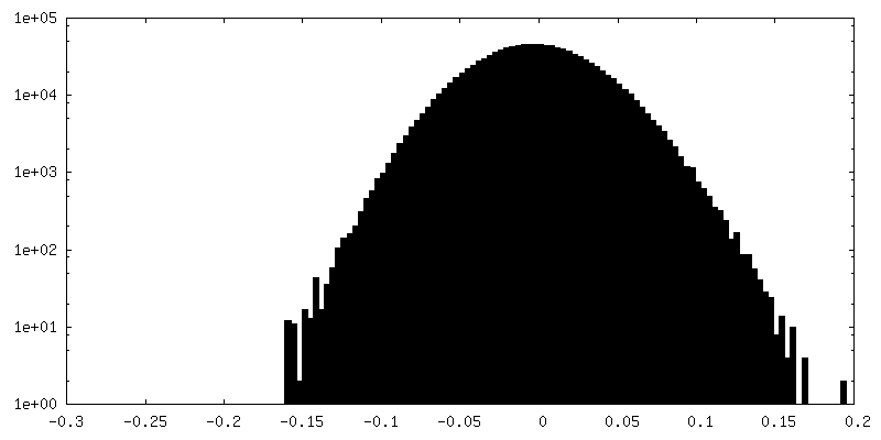
| Density Histograms |

-Half map: #1
| File | emd_4517_half_map_2.map | ||||||||||||
|---|---|---|---|---|---|---|---|---|---|---|---|---|---|
| Projections & Slices |
| ||||||||||||
| Density Histograms |
- Sample components
-Entire : MloK1 potassium channel in lipidic bilayer of 2D crystals
| Entire | Name: MloK1 potassium channel in lipidic bilayer of 2D crystals |
|---|---|
| Components |
|
-Supramolecule #1: MloK1 potassium channel in lipidic bilayer of 2D crystals
| Supramolecule | Name: MloK1 potassium channel in lipidic bilayer of 2D crystals type: complex / ID: 1 / Parent: 0 / Macromolecule list: #1 |
|---|---|
| Source (natural) | Organism: Mesorhizobium loti MAFF303099 (bacteria) |
| Molecular weight | Theoretical: 160 KDa |
-Macromolecule #1: Cyclic nucleotide-gated potassium channel mll3241
| Macromolecule | Name: Cyclic nucleotide-gated potassium channel mll3241 / type: protein_or_peptide / ID: 1 / Number of copies: 4 / Enantiomer: LEVO |
|---|---|
| Source (natural) | Organism: Mesorhizobium loti MAFF303099 (bacteria) |
| Molecular weight | Theoretical: 37.766297 KDa |
| Recombinant expression | Organism: |
| Sequence | String: MSVLPFLRIY APLNAVLAAP GLLAVAALTI PDMSGRSRLA LAALLAVIWG AYLLQLAATL LKRRAGVVRD RTPKIAIDVL AVLVPLAAF LLDGSPDWSL YCAVWLLKPL RDSTFFPVLG RVLANEARNL IGVTTLFGVV LFAVALAAYV IERDIQPEKF G SIPQAMWW ...String: MSVLPFLRIY APLNAVLAAP GLLAVAALTI PDMSGRSRLA LAALLAVIWG AYLLQLAATL LKRRAGVVRD RTPKIAIDVL AVLVPLAAF LLDGSPDWSL YCAVWLLKPL RDSTFFPVLG RVLANEARNL IGVTTLFGVV LFAVALAAYV IERDIQPEKF G SIPQAMWW AVVTLSTTGY GDTIPQSFAG RVLAGAVMMS GIGIFGLWAG ILATGFYQEV RRGDFVRNWQ LVAAVPLFQK LG PAVLVEI VRALRARTVP AGAVICRIGE PGDRMFFVVE GSVSVATPNP VELGPGAFFG EMALISGEPR SATVSAATTV SLL SLHSAD FQMLCSSSPE IAEIFRKTAL ERRGAAASA UniProtKB: Cyclic nucleotide-gated potassium channel mll3241 |
-Macromolecule #2: POTASSIUM ION
| Macromolecule | Name: POTASSIUM ION / type: ligand / ID: 2 / Number of copies: 2 / Formula: K |
|---|---|
| Molecular weight | Theoretical: 39.098 Da |
-Experimental details
-Structure determination
| Method | cryo EM |
|---|---|
 Processing Processing | single particle reconstruction |
| Aggregation state | 2D array |
-Sample preparation
| Buffer | pH: 7.6 Details: 20 mM KCl, 20 mM Tris-HCl pH 7.6, 1 mM BaCl2, 1 mM EDTA, 0.2 mM cAMP |
|---|---|
| Vitrification | Cryogen name: ETHANE |
- Electron microscopy
| Microscope | FEI TITAN KRIOS |
|---|---|
| Image recording | Film or detector model: GATAN K2 SUMMIT (4k x 4k) / Detector mode: COUNTING / Number real images: 270 / Average exposure time: 16.0 sec. / Average electron dose: 45.0 e/Å2 Details: The dataset of 346 movies recorded and processed for Kowal et al. (2018) 2 was employed. As reported, the data were collected on an FEI Titan Krios TEM equipped with a Gatan K2 DED. Total ...Details: The dataset of 346 movies recorded and processed for Kowal et al. (2018) 2 was employed. As reported, the data were collected on an FEI Titan Krios TEM equipped with a Gatan K2 DED. Total dose: 40 e-/A^2 distributed over 40 movie frames. Pixel size: 1.3 A on the sample level (counting mode). Nominal tilt range: -55 to +55 degrees. |
| Electron beam | Acceleration voltage: 300 kV / Electron source:  FIELD EMISSION GUN FIELD EMISSION GUN |
| Electron optics | Illumination mode: OTHER / Imaging mode: BRIGHT FIELD / Cs: 2.7 mm / Nominal defocus max: 4.3 µm / Nominal defocus min: 0.75 µm / Nominal magnification: 50000 |
| Sample stage | Specimen holder model: FEI TITAN KRIOS AUTOGRID HOLDER / Cooling holder cryogen: NITROGEN |
| Experimental equipment |  Model: Titan Krios / Image courtesy: FEI Company |
+Image processing
-Atomic model buiding 1
| Initial model | PDB ID: Chain - Source name: PDB / Chain - Initial model type: experimental model |
|---|---|
| Details | The consensus model was flexibily fit in to the EM map of this class using Normal Mode Analysis with iMODFIT, and then real-space refined using PHENIX. |
| Refinement | Protocol: FLEXIBLE FIT |
| Output model | PDB-6qd2: |
