Movie
Movie Controller

[English] 日本語
 Yorodumi
- EMDB-19906: In situ nucleosome subtomogram average from Chlamydomonas reinhardtii -
+ Open data

- Basic information
| Entry |  | |||||||||
|---|---|---|---|---|---|---|---|---|---|---|
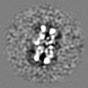
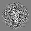
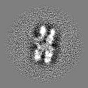
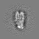
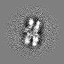
| Title | In situ nucleosome subtomogram average from Chlamydomonas reinhardtii | |||||||||
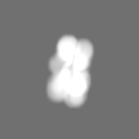
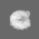
 Map data Map data | ||||||||||
 Sample Sample |
| |||||||||
 Keywords Keywords | nucleosome / histone / cryo-et / chlamydomonas / chromatin / tomography / dna / in situ | |||||||||
| Biological species |   Chlamydomonas reinhardtii (plant) Chlamydomonas reinhardtii (plant) | |||||||||
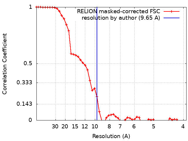
| Method | subtomogram averaging / cryo EM / Resolution: 9.65 Å | |||||||||
 Authors Authors | Michael AK / Righetto RD / Obr M / van der Stappen P / Lamm L / Khavnekar S / Kotecha A / Engel BD | |||||||||
| Funding support |  Switzerland, 1 items Switzerland, 1 items
| |||||||||
 Citation Citation | Journal: Mol Cell / Year: 2026 Title: Toward community-driven visual proteomics with large-scale cryo-electron tomography of Chlamydomonas reinhardtii. Authors: Ron Kelley / Sagar Khavnekar / Ricardo D Righetto / Jessica Heebner / Martin Obr / Xianjun Zhang / Saikat Chakraborty / Grigory Tagiltsev / Alicia K Michael / Sofie van Dorst / Florent Waltz ...Authors: Ron Kelley / Sagar Khavnekar / Ricardo D Righetto / Jessica Heebner / Martin Obr / Xianjun Zhang / Saikat Chakraborty / Grigory Tagiltsev / Alicia K Michael / Sofie van Dorst / Florent Waltz / Caitlyn L McCafferty / Lorenz Lamm / Simon Zufferey / Philippe Van der Stappen / Hugo van den Hoek / Wojciech Wietrzynski / Pavol Harar / William Wan / John A G Briggs / Jürgen M Plitzko / Benjamin D Engel / Abhay Kotecha /     Abstract: In situ cryo-electron tomography (cryo-ET) has emerged as the method of choice to investigate the structures of biomolecules in their native context. However, challenges remain for the efficient ...In situ cryo-electron tomography (cryo-ET) has emerged as the method of choice to investigate the structures of biomolecules in their native context. However, challenges remain for the efficient production and sharing of large-scale cryo-ET datasets. Here, we combined cryogenic plasma-based focused ion beam (cryo-PFIB) milling with recent advances in cryo-ET acquisition and processing to generate a dataset of 1,829 annotated tomograms of the green alga Chlamydomonas reinhardtii, which we provide as a community resource to drive method development and inspire biological discovery. To assay data quality, we performed subtomogram averaging of both soluble and membrane-bound complexes ranging in size from >3 MDa to ∼200 kDa, including 80S ribosomes, Rubisco, nucleosomes, microtubules, clathrin, photosystem II, and mitochondrial ATP synthase. The majority of these density maps reached sub-nanometer resolution, demonstrating the potential of this C. reinhardtii dataset as well as the promise of modern cryo-ET workflows and open data sharing to empower visual proteomics. #1: Journal: Biorxiv / Year: 2024Title: Towards community-driven visual proteomics with large-scale cryo-electron tomography of Chlamydomonas reinhardtii Authors: Kelley R / Khavnekar S / Righetto RD / Heebner J / Obr M / Zhang X / Chakraborty S / Tagiltsev G / Michael AK / van Dorst S / Waltz F / McCafferty CL / Lamm L / Zufferey S / Van der Stappen ...Authors: Kelley R / Khavnekar S / Righetto RD / Heebner J / Obr M / Zhang X / Chakraborty S / Tagiltsev G / Michael AK / van Dorst S / Waltz F / McCafferty CL / Lamm L / Zufferey S / Van der Stappen P / van den Hoek H / Wietrzynski W / Harar P / Wan W / Briggs JA / Plitzko JM / Engel BD / Kotecha A | |||||||||
| History |
|
- Structure visualization
| Supplemental images |
|---|

- Downloads & links
-EMDB archive
| Map data | emd_19906.map.gz | 7.5 MB |  EMDB map data format EMDB map data format | |
|---|---|---|---|---|
| Header (meta data) | emd-19906-v30.xmlemd-19906.xml | 19.1 KB 19.1 KB | Display Display | EMDB header |
| FSC (resolution estimation) | emd_19906_fsc.xml | 4.7 KB | Display | FSC data file |
| Images | 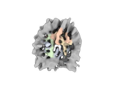 emd_19906.png emd_19906.png | 42.8 KB | ||
| Masks | emd_19906_msk_1.map | 8 MB | Mask map | |
| Filedesc metadata | emd-19906.cif.gz | 4.9 KB | ||
| Others | emd_19906_half_map_1.map.gzemd_19906_half_map_2.map.gz | 4 MB 4 MB | ||
| Archive directory |  http://ftp.pdbj.org/pub/emdb/structures/EMD-19906ftp://ftp.pdbj.org/pub/emdb/structures/EMD-19906 http://ftp.pdbj.org/pub/emdb/structures/EMD-19906ftp://ftp.pdbj.org/pub/emdb/structures/EMD-19906 | HTTPS FTP |
-Related structure data


-Links
| EMDB pages | EMDB (EBI/PDBe) / EMDataResource |
|---|
-Map
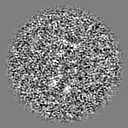
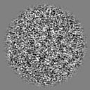
| File | Download / File: emd_19906.map.gz / Format: CCP4 / Size: 8 MB / Type: IMAGE STORED AS FLOATING POINT NUMBER (4 BYTES) | ||||||||||||||||||||||||||||||||||||
|---|---|---|---|---|---|---|---|---|---|---|---|---|---|---|---|---|---|---|---|---|---|---|---|---|---|---|---|---|---|---|---|---|---|---|---|---|---|
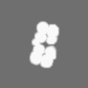
| Projections & slices | Image control
Images are generated by Spider. | ||||||||||||||||||||||||||||||||||||
| Voxel size | X=Y=Z: 1.96 Å | ||||||||||||||||||||||||||||||||||||
| Density |
| ||||||||||||||||||||||||||||||||||||
| Symmetry | Space group: 1 | ||||||||||||||||||||||||||||||||||||
| Details | EMDB XML:
|
 Z (Sec.)
 Y (Row.)
 X (Col.)


-Supplemental data
-Mask #1
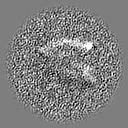
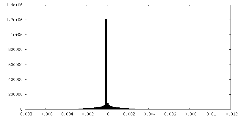
| File | emd_19906_msk_1.map | ||||||||||||
|---|---|---|---|---|---|---|---|---|---|---|---|---|---|
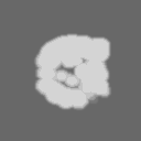
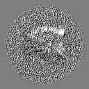
| Projections & Slices |
| ||||||||||||
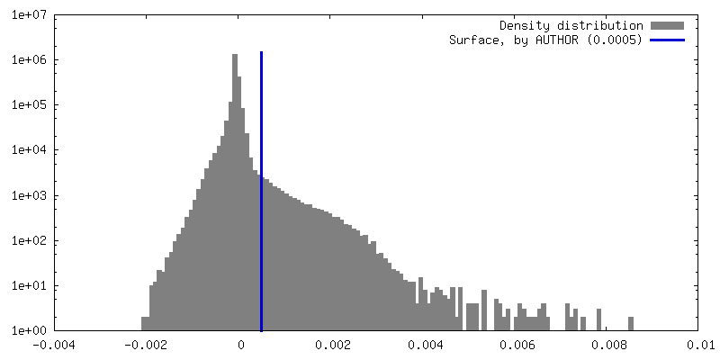
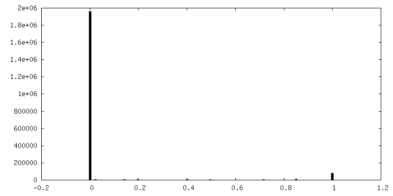
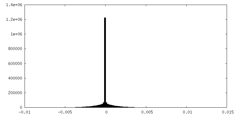
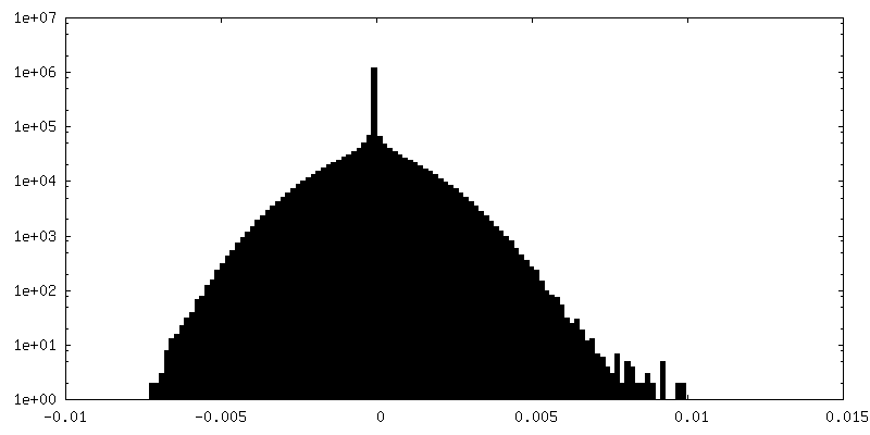
| Density Histograms |


-Half map: #1
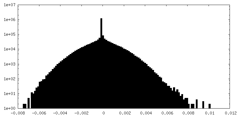
| File | emd_19906_half_map_1.map | ||||||||||||
|---|---|---|---|---|---|---|---|---|---|---|---|---|---|
| Projections & Slices |
| ||||||||||||
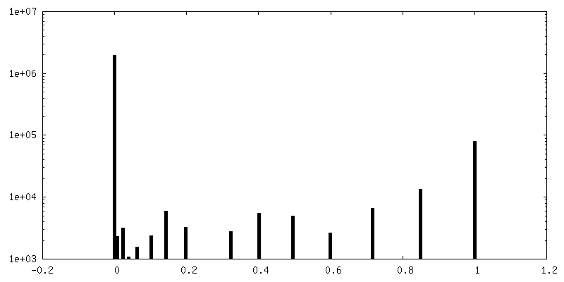
| Density Histograms |
-Half map: #2
| File | emd_19906_half_map_2.map | ||||||||||||
|---|---|---|---|---|---|---|---|---|---|---|---|---|---|
| Projections & Slices |
| ||||||||||||
| Density Histograms |
- Sample components
-Entire : Chlamydomonas reinhardtii
| Entire | Name: Chlamydomonas reinhardtii (plant) |
|---|---|
| Components |
|
-Supramolecule #1: Chlamydomonas reinhardtii
| Supramolecule | Name: Chlamydomonas reinhardtii / type: cell / ID: 1 / Parent: 0 |
|---|---|
| Source (natural) | Organism: Chlamydomonas reinhardtii (plant) / Strain: CC-3994 mat3-4 mt+ |
-Experimental details
-Structure determination
| Method | cryo EM |
|---|---|
 Processing Processing | subtomogram averaging |
| Aggregation state | cell |
-Sample preparation
| Buffer | pH: 7 |
|---|---|
| Grid | Model: Quantifoil R2/1 / Material: COPPER / Pretreatment - Type: PLASMA CLEANING |
| Vitrification | Cryogen name: ETHANE-PROPANE / Instrument: FEI VITROBOT MARK IV |
- Electron microscopy
| Microscope | TFS KRIOS |
|---|---|
| Specialist optics | Energy filter - Name: TFS Selectris X / Energy filter - Slit width: 10 eV |
| Image recording | Film or detector model: TFS FALCON 4i (4k x 4k) / Digitization - Dimensions - Width: 4096 pixel / Digitization - Dimensions - Height: 4096 pixel / Average electron dose: 3.5 e/Å2 |
| Electron beam | Acceleration voltage: 300 kV / Electron source:  FIELD EMISSION GUN FIELD EMISSION GUN |
| Electron optics | Illumination mode: FLOOD BEAM / Imaging mode: BRIGHT FIELD / Nominal defocus max: 3.5 µm / Nominal defocus min: 0.8 µm / Nominal magnification: 64000 |
| Sample stage | Specimen holder model: FEI TITAN KRIOS AUTOGRID HOLDER / Cooling holder cryogen: NITROGEN |
| Experimental equipment |  Model: Titan Krios / Image courtesy: FEI Company |