 ムービー
ムービー コントローラー
 構造ビューア
 EMN検索について


-検索条件
-検索結果
検索 (著者・登録者: nickel & l)の結果86件中、1から50件目までを表示しています

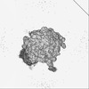
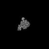
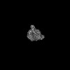
EMDB-72553: 
Escherichia coli transcription-translation loosely coupled complex (TTC-LC^walked) containing mRNA with a 39 nt long spacer, NusG, NusA, and fMet-tRNAs in E-site and P-site - Map 1a
手法: 単粒子 / : Shandilya S, Molodtsov V, Wang C, Ebright RH

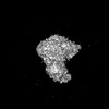
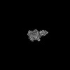
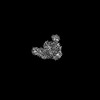
EMDB-72554:
Escherichia coli transcription-translation loosely coupled complex complex (TTC-LC^walked) containing mRNA with a 39 nt long spacer, NusG, NusA, and fMet-tRNAs in E-site and P-site - Map 1
手法: 単粒子 / : Shandilya S, Molodtsov V, Wang C, Ebright RH

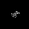
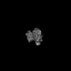
EMDB-72646:
Escherichia coli transcription-translation loosely coupled complex (TTC-LC^walked) containing mRNA with a 39 nt long spacer, NusG, NusA, and fMet-tRNAs in E-site and P-site
手法: 単粒子 / : Shandilya S, Wang C, Molodtsov V, Ebright RH

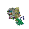
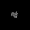
PDB-9y79:
Escherichia coli transcription-translation loosely coupled complex (TTC-LC^walked) containing mRNA with a 39 nt long spacer, NusG, NusA, and fMet-tRNAs in E-site and P-site
手法: 単粒子 / : Shandilya S, Wang C, Molodtsov V, Ebright RH

EMDB-43332:
Escherichia coli transcription-translation coupled complex (TTC-B) containing mRNA with a 33 nt long spacer, ops signal, RfaH, NusA, and fMet-tRNAs in E-site and P-site
手法: 単粒子 / : Molodtsov V, Wang C, Ebright RH

EMDB-43335:
Escherichia coli transcription-translation loosely coupled complex (TTC-LC) containing mRNA with a 36 nt long spacer, ops signal, RfaH, NusA, and fMet-tRNAs in E-site and P-site
手法: 単粒子 / : Molodtsov V, Wang C, Ebright RH
EMDB-43387:
Escherichia coli transcription-translation loosely coupled complex (TTC-LC) containing mRNA with a 39 nt long spacer, ops signal, RfaH, NusA, and fMet-tRNAs in E-site and P-site
手法: 単粒子 / : Molodtsov V, Wang C, Ebright RH

EMDB-43388:
Escherichia coli transcription-translation coupled complex (TTC-B) containing mRNA with a 36 nt long spacer, NusG, NusA, and fMet-tRNAs in E-site and P-site
手法: 単粒子 / : Molodtsov V, Wang C, Ebright RH

EMDB-43389:
Escherichia coli transcription-translation loosely coupled complex (TTC-LC) containing mRNA with a 39 nt long spacer, NusG, NusA, and fMet-tRNAs in E-site and P-site
手法: 単粒子 / : Molodtsov V, Wang C, Ebright RH

EMDB-43390:
Escherichia coli transcription-translation loosely coupled complex (TTC-LC) containing mRNA with a 51 nt long spacer, NusG, NusA, and fMet-tRNAs in E-site and P-site
手法: 単粒子 / : Molodtsov V, Wang C, Ebright RH

EMDB-43391:
Escherichia coli transcription-translation loosely coupled complex (TTC-LC) containing mRNA with a 60 nt long spacer, NusG, NusA, and fMet-tRNAs in E-site and P-site
手法: 単粒子 / : Molodtsov V, Wang C, Ebright RH

PDB-8vkv:
Escherichia coli transcription-translation coupled complex (TTC-B) containing mRNA with a 33 nt long spacer, ops signal, RfaH, NusA, and fMet-tRNAs in E-site and P-site
手法: 単粒子 / : Molodtsov V, Wang C, Ebright RH

PDB-8vl1:
Escherichia coli transcription-translation loosely coupled complex (TTC-LC) containing mRNA with a 36 nt long spacer, ops signal, RfaH, NusA, and fMet-tRNAs in E-site and P-site
手法: 単粒子 / : Molodtsov V, Wang C, Ebright RH

PDB-8voo:
Escherichia coli transcription-translation loosely coupled complex (TTC-LC) containing mRNA with a 39 nt long spacer, ops signal, RfaH, NusA, and fMet-tRNAs in E-site and P-site
手法: 単粒子 / : Molodtsov V, Wang C, Ebright RH

PDB-8vop:
Escherichia coli transcription-translation coupled complex (TTC-B) containing mRNA with a 36 nt long spacer, NusG, NusA, and fMet-tRNAs in E-site and P-site
手法: 単粒子 / : Molodtsov V, Wang C, Ebright RH
PDB-8voq:
Escherichia coli transcription-translation loosely coupled complex (TTC-LC) containing mRNA with a 39 nt long spacer, NusG, NusA, and fMet-tRNAs in E-site and P-site
手法: 単粒子 / : Molodtsov V, Wang C, Ebright RH

PDB-8vor:
Escherichia coli transcription-translation loosely coupled complex (TTC-LC) containing mRNA with a 51 nt long spacer, NusG, NusA, and fMet-tRNAs in E-site and P-site
手法: 単粒子 / : Molodtsov V, Wang C, Ebright RH
PDB-8vos:
Escherichia coli transcription-translation loosely coupled complex (TTC-LC) containing mRNA with a 60 nt long spacer, NusG, NusA, and fMet-tRNAs in E-site and P-site
手法: 単粒子 / : Molodtsov V, Wang C, Ebright RH

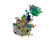
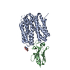
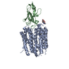
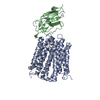
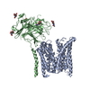
EMDB-53511:
SpCas9 with computationally designed SpCas9_b10 binder
手法: 単粒子 / : Pacesa M, Nickel L, Correia BE
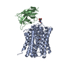
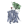
EMDB-53510:
SpCas9 with computationally designed SpCas9_b3 binder
手法: 単粒子 / : Pacesa M, Nickel L, Correia BE
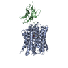
EMDB-19716:
Cryo-EM structure of apo human SLC19A3 in outward-open state
手法: 単粒子 / : Gabriel F, Loew C
EMDB-19750:
Cryo-EM structure of thiamine-bound human SLC19A3 in outward-open state
手法: 単粒子 / : Gabriel F, Loew C
EMDB-19752:
Cryo-EM structure of fedratinib-bound human SLC19A3 in inward-open state
手法: 単粒子 / : Gabriel F, Loew C
EMDB-19753:
Cryo-EM structure of hydroxychloroquine-bound human SLC19A3 in inward-open state
手法: 単粒子 / : Gabriel F, Loew C
EMDB-19754:
Cryo-EM structure of thiamine-bound human SLC19A3 in inward-open state
手法: 単粒子 / : Gabriel F, Loew C
EMDB-19755:
Cryo-EM structure of amprolium-bound human SLC19A3 in inward-open state
手法: 単粒子 / : Gabriel F, Loew C
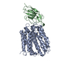
EMDB-51088:
Cryo-EM structure of apo human SLC19A3 in inward-open state
手法: 単粒子 / : Gabriel F, Loew C
PDB-8s5u:
Cryo-EM structure of thiamine-bound human SLC19A3 in outward-open state
手法: 単粒子 / : Gabriel F, Loew C
PDB-8s5w:
Cryo-EM structure of fedratinib-bound human SLC19A3 in inward-open state
手法: 単粒子 / : Gabriel F, Loew C
PDB-8s5z:
Cryo-EM structure of hydroxychloroquine-bound human SLC19A3 in inward-open state
手法: 単粒子 / : Gabriel F, Loew C
PDB-8s61:
Cryo-EM structure of thiamine-bound human SLC19A3 in inward-open state
手法: 単粒子 / : Gabriel F, Loew C
PDB-8s62:
Cryo-EM structure of amprolium-bound human SLC19A3 in inward-open state
手法: 単粒子 / : Gabriel F, Loew C

EMDB-19005:
structure of the GLMP/MFSD1 complex
手法: 単粒子 / : Jungnickel KEJ, Loew C
EMDB-19006:
Lysosomal peptide transporter
手法: 単粒子 / : Jungnickel KEJ, Loew C
PDB-8r8q:
Lysosomal peptide transporter
手法: 単粒子 / : Jungnickel KEJ, Loew C

EMDB-15244:
Tomogram of an Ebola VLP composed of GP, VP40, NP, VP24 and VP35 at pH 7.4 (Figure 1A-D)
手法: トモグラフィー / : Winter SL, Chlanda P

EMDB-15268:
Tomogram of an Ebola VLP composed of VP40 at pH 4.5 (Figure 1J)
手法: トモグラフィー / : Winter SL, Chlanda P
EMDB-15951:
Tomogram of an EBOV-infected Huh7 cell showing a late endosome with internalized EBOV particles
手法: トモグラフィー / : Winter SL, Chlanda P

EMDB-15956:
Tomogram of an extracellular EBOV particle adjacent to an EBOV-infected Huh7 cell
手法: トモグラフィー / : Winter SL, Chlanda P

EMDB-16128:
Tomogram of a late endosome of A549 cell infected with influenza A virus.
手法: トモグラフィー / : Klein S, Chlanda P

EMDB-16129:
Tomogram of a late endosome of A549 cell infected with influenza A virus (Figure 6C).
手法: トモグラフィー / : Klein S, Chlanda P

EMDB-16130:
Tomogram of a late endosome of A549 cell infected with influenza A virus (Figure 6E)
手法: トモグラフィー / : Klein S, Chlanda P

EMDB-16131:
Tomogram of a late endosome of A549 cell infected with influenza A virus (Figure 6G)
手法: トモグラフィー / : Klein S, Chlanda P

EMDB-16132:
Tomogram of a late endosome of A549 cell infected with influenza A virus (Figure 6I,K,M)
手法: トモグラフィー / : Klein S, Chlanda P

EMDB-16133:
Tomogram of a late endosome of A549 cell infected with influenza A virus (Figure 6O)
手法: トモグラフィー / : Klein S, Chlanda P
EMDB-15705:
Tomogram of a late endosome of A549 cell (Figure 1R)
手法: トモグラフィー / : Klein S, Chlanda P
EMDB-15707:
Tomogram of a late endosome of A549 cell treated with IFN-beta (Figure 1T)
手法: トモグラフィー / : Klein S, Chlanda P
EMDB-15708:
Tomogram of a late endosome of A549-IFITM3 cell (Figure 1V)
手法: トモグラフィー / : Klein S, Chlanda P
ページ:
 wwPDBはEMDBデータモデルのバージョン3へ移行します
