 Movie
Movie Controller


+ Open data

- Basic information
| Entry | Database: PDB / ID: 8s4u | ||||||
|---|---|---|---|---|---|---|---|
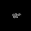
| Title | Cryo-EM structure of apo human SLC19A3 in outward-open state | ||||||
 Components Components |
| ||||||
 Keywords Keywords | MEMBRANE PROTEIN / SLC19A3 / vitamin transporter / thiamine transporter / MFS fold / nanobody complex | ||||||
| Function / homology |  Function and homology information Function and homology informationpyridoxine transport / thiamine-containing compound metabolic process / Vitamin B1 (thiamin) metabolism / thiamine transmembrane transport / thiamine transmembrane transporter activity / thiamine transport / thiamine diphosphate biosynthetic process / membrane / plasma membrane Similarity search - Function | ||||||
| Biological species |   Homo sapiens (human) Homo sapiens (human) | ||||||
| Method | ELECTRON MICROSCOPY / single particle reconstruction / cryo EM / Resolution: 3.09 Å | ||||||
 Authors Authors | Gabriel, F. / Loew, C. | ||||||
| Funding support | European Union, 1items
| ||||||
 Citation Citation | Journal: Nat Commun / Year: 2024 Title: Structural basis of thiamine transport and drug recognition by SLC19A3. Authors: Florian Gabriel / Lea Spriestersbach / Antonia Fuhrmann / Katharina E J Jungnickel / Siavash Mostafavi / Els Pardon / Jan Steyaert / Christian Löw /   Abstract: Thiamine (vitamin B) functions as an essential coenzyme in cells. Humans and other mammals cannot synthesise this vitamin de novo and thus have to take it up from their diet. Eventually, every cell ...Thiamine (vitamin B) functions as an essential coenzyme in cells. Humans and other mammals cannot synthesise this vitamin de novo and thus have to take it up from their diet. Eventually, every cell needs to import thiamine across its plasma membrane, which is mainly mediated by the two specific thiamine transporters SLC19A2 and SLC19A3. Loss of function mutations in either of these transporters lead to detrimental, life-threatening metabolic disorders. SLC19A3 is furthermore a major site of drug interactions. Many medications, including antidepressants, antibiotics and chemotherapeutics are known to inhibit this transporter, with potentially fatal consequences for patients. Despite a thorough functional characterisation over the past two decades, the structural basis of its transport mechanism and drug interactions has remained elusive. Here, we report seven cryo-electron microscopy (cryo-EM) structures of the human thiamine transporter SLC19A3 in complex with various ligands. Conformation-specific nanobodies enable us to capture different states of SLC19A3's transport cycle, revealing the molecular details of thiamine recognition and transport. We identify seven previously unknown drug interactions of SLC19A3 and present structures of the transporter in complex with the inhibitors fedratinib, amprolium and hydroxychloroquine. These data allow us to develop an understanding of the transport mechanism and ligand recognition of SLC19A3. | ||||||
| History |
|
- Structure visualization
| Structure viewer | Molecule: MolmilJmol/JSmol |
|---|
- Downloads & links
-Download
| PDBx/mmCIF format | 8s4u.cif.gz | 103.2 KB | Display | PDBx/mmCIF format |
|---|---|---|---|---|
| PDB format | pdb8s4u.ent.gz | 71.5 KB | Display | PDB format |
| PDBx/mmJSON format | 8s4u.json.gz | Tree view | PDBx/mmJSON format | |
| Others |  Other downloads Other downloads |
-Validation report
| Arichive directory | https://data.pdbj.org/pub/pdb/validation_reports/s4/8s4uftp://data.pdbj.org/pub/pdb/validation_reports/s4/8s4u | HTTPS FTP |
|---|
-Related structure data
| Related structure data |  19716MC 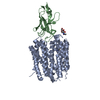 8s5uC 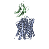 8s5wC 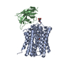 8s5zC 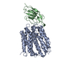 8s61C 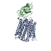 8s62C 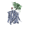 9g5kC M: map data used to model this data C: citing same article ( |
|---|---|
| Similar structure data |
-Links
 PDBj
PDBj


- Assembly
| Deposited unit | 
|
|---|---|
| 1 |
|
-Components
| #1: Antibody | Mass: 15217.906 Da / Num. of mol.: 1 Source method: isolated from a genetically manipulated source Source: (gene. exp.)  |
|---|---|
| #2: Protein | Mass: 59687.609 Da / Num. of mol.: 1 Source method: isolated from a genetically manipulated source Source: (gene. exp.) Homo sapiens (human) / Gene: SLC19A3 / Production host: Homo sapiens (human) / References: UniProt: Q9BZV2 |
| #3: Sugar | ChemComp-NAG /   Type: D-saccharide, beta linking / Mass: 221.208 Da / Num. of mol.: 1 / Source method: obtained synthetically / Formula: C8H15NO6 Type: D-saccharide, beta linking / Mass: 221.208 Da / Num. of mol.: 1 / Source method: obtained synthetically / Formula: C8H15NO6 |
| Has ligand of interest | N |
| Has protein modification | Y |
-Experimental details
-Experiment
| Experiment | Method: ELECTRON MICROSCOPY |
|---|---|
| EM experiment | Aggregation state: PARTICLE / 3D reconstruction method: single particle reconstruction |
- Sample preparation
| Component |
| |||||||||||||||||||||||||
|---|---|---|---|---|---|---|---|---|---|---|---|---|---|---|---|---|---|---|---|---|---|---|---|---|---|---|
| Molecular weight | Value: 0.075 MDa / Experimental value: NO | |||||||||||||||||||||||||
| Source (natural) |
| |||||||||||||||||||||||||
| Source (recombinant) |
| |||||||||||||||||||||||||
| Buffer solution | pH: 7.4 | |||||||||||||||||||||||||
| Buffer component |
| |||||||||||||||||||||||||
| Specimen | Embedding applied: NO / Shadowing applied: NO / Staining applied: NO / Vitrification applied: YES | |||||||||||||||||||||||||
| Vitrification | Instrument: FEI VITROBOT MARK IV / Cryogen name: ETHANE-PROPANE / Humidity: 100 % / Chamber temperature: 283.15 K |
- Electron microscopy imaging
| Experimental equipment |  Model: Titan Krios / Image courtesy: FEI Company |
|---|---|
| Microscopy | Model: FEI TITAN KRIOS |
| Electron gun | Electron source:  FIELD EMISSION GUN / Accelerating voltage: 300 kV / Illumination mode: SPOT SCAN FIELD EMISSION GUN / Accelerating voltage: 300 kV / Illumination mode: SPOT SCAN |
| Electron lens | Mode: BRIGHT FIELD / Nominal magnification: 105000 X / Nominal defocus max: 1600 nm / Nominal defocus min: 800 nm / Cs: 2.7 mm / C2 aperture diameter: 70 µm |
| Specimen holder | Cryogen: NITROGEN |
| Image recording | Electron dose: 45 e/Å2 / Film or detector model: GATAN K3 BIOQUANTUM (6k x 4k) / Num. of grids imaged: 1 / Num. of real images: 5859 |
| EM imaging optics | Energyfilter name: GIF Bioquantum / Energyfilter slit width: 20 eV |
- Processing
| EM software |
| |||||||||||||||||||||||||||||||||||||||||||||||||||||||||||||||||||||||||||||||||||||||||||||||||||||||||||
|---|---|---|---|---|---|---|---|---|---|---|---|---|---|---|---|---|---|---|---|---|---|---|---|---|---|---|---|---|---|---|---|---|---|---|---|---|---|---|---|---|---|---|---|---|---|---|---|---|---|---|---|---|---|---|---|---|---|---|---|---|---|---|---|---|---|---|---|---|---|---|---|---|---|---|---|---|---|---|---|---|---|---|---|---|---|---|---|---|---|---|---|---|---|---|---|---|---|---|---|---|---|---|---|---|---|---|---|---|
| CTF correction | Type: PHASE FLIPPING AND AMPLITUDE CORRECTION | |||||||||||||||||||||||||||||||||||||||||||||||||||||||||||||||||||||||||||||||||||||||||||||||||||||||||||
| 3D reconstruction | Resolution: 3.09 Å / Resolution method: FSC 0.143 CUT-OFF / Num. of particles: 486079 / Symmetry type: POINT | |||||||||||||||||||||||||||||||||||||||||||||||||||||||||||||||||||||||||||||||||||||||||||||||||||||||||||
| Atomic model building | Protocol: AB INITIO MODEL / Space: REAL | |||||||||||||||||||||||||||||||||||||||||||||||||||||||||||||||||||||||||||||||||||||||||||||||||||||||||||
| Atomic model building | Source name: AlphaFold / Type: in silico model | |||||||||||||||||||||||||||||||||||||||||||||||||||||||||||||||||||||||||||||||||||||||||||||||||||||||||||
| Refinement | Resolution: 3.09→3.09 Å / Cor.coef. Fo:Fc: 0.685 / WRfactor Rwork: 0.392 / Average fsc free: 0 / Average fsc overall: 0.8037 / Average fsc work: 0.8037 / ESU R: 0.625 Details: Hydrogens have been added in their riding positions
| |||||||||||||||||||||||||||||||||||||||||||||||||||||||||||||||||||||||||||||||||||||||||||||||||||||||||||
| Solvent computation | Solvent model: NONE | |||||||||||||||||||||||||||||||||||||||||||||||||||||||||||||||||||||||||||||||||||||||||||||||||||||||||||
| Displacement parameters | Biso mean: 39.382 Å2 | |||||||||||||||||||||||||||||||||||||||||||||||||||||||||||||||||||||||||||||||||||||||||||||||||||||||||||
| Refine LS restraints |
|