 ムービー
ムービー コントローラー
 構造ビューア
 EMN検索について


-検索条件
-検索結果
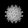
検索 (著者・登録者: mosalaganti & s)の結果70件中、1から50件目までを表示しています
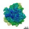
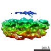
EMDB-42943: 
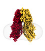
Structure of the human systemic RNAi defective transmembrane protein 1 (hSIDT1)
PDB-8v38:
Structure of the human systemic RNAi defective transmembrane protein 1 (hSIDT1)
EMDB-41620:
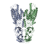
Cryo-EM structure of HGSNAT-acetyl-CoA complex at pH 7.5
PDB-8tu9:
Cryo-EM structure of HGSNAT-acetyl-CoA complex at pH 7.5

EMDB-43271:
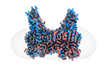
Cryo-EM structure of the Helicobacter pylori VacA hexamer that was detergent solubilized from membrane, C6 symmetry applied

EMDB-43272:
Cryo-EM structure of Helicobacter pylori VacA hexamer that was detergent solubilized form membrane, no symmetry applied

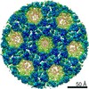
EMDB-16772:
Subtomogram average of Immature Rotavirus TLP penton
EMDB-16773:
In situ STA of rotavirus TLP (icos)

PDB-8co6:
Subtomogram average of Immature Rotavirus TLP penton
EMDB-16762:
In situ map of Rotavirus SLP

EMDB-16146:
SPA of Trypsin untreated Rotavirus TLP spike

EMDB-16769:
In situ STA of rotavirus DLP (penton)

EMDB-16771:
In situ STA of Rotavirus enveloped DLP (icos)
EMDB-16774:
in situ Subtomogram average of Immature Rotavirus TLP spike

PDB-8bp8:
SPA of Trypsin untreated Rotavirus TLP spike

PDB-8coa:
in situ Subtomogram average of Immature Rotavirus TLP spike
EMDB-16767:
In situ STA of rotavirus enveloped DLP (penton)

EMDB-25804:
Cryo-EM structure of methane monooxygenase hydroxylase (by quantifoil)

EMDB-25805:
Cryo-EM structure of methane monooxygenase hydroxylase (by graphene)

PDB-7tc7:
Cryo-EM structure of methane monooxygenase hydroxylase (by quantifoil)

PDB-7tc8:
Cryo-EM structure of methane monooxygenase hydroxylase (by graphene)

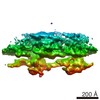
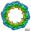
EMDB-14325:
Cytoplasmic ring of the human nuclear pore complex from isolated HeLa nuclear envelopes

EMDB-14326:
Structure of nuclear pore complex from HEK cells with GP210 knockout

EMDB-14327:
Nuclear pore complex from intact HeLa cells

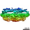
EMDB-14328:
Inner/spoke ring of the human nuclear pore complex from isolated HeLa nuclear envelopes.

EMDB-14330:
Nuclear ring of the human nuclear pore complex from isolated HeLa nuclear envelopes

EMDB-14321:
Human nuclear pore complex (dilated)
PDB-7r5j:
Human nuclear pore complex (dilated)

EMDB-14322:
Human nuclear pore complex (constricted)
PDB-7r5k:
Human nuclear pore complex (constricted)

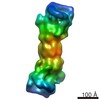
EMDB-14243:
cryoEM structure of human Nup155 (residues 19-981)

PDB-7r1y:
cryoEM structure of human Nup155 (residues 19-981)

EMDB-11222:
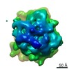
Structure of SARS-CoV-2 spike glycoprotein (S) trimer determined by sub-tomogram averaging

EMDB-11223:
Structure of SARS-CoV-2 spike glycoprotein (S) monomer in a closed conformation determined by sub-tomogram averaging
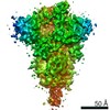
EMDB-11347:
Structure of SARS-CoV-2 spike glycoprotein (S) trimer with one receptor binding domain (RBD) in open-state determined by subtomogram averaging
EMDB-22190:
Subtomogram averaging map of yeast polysome
EMDB-22196:
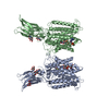
Cryo-EM Structure of K63R Ubiquitin Mutant Ribosome under Oxidative Stress

EMDB-22197:
Cryo-EM Structure of K63R Ubiquitin Mutant Consensus Ribosome under Oxidative Stress

EMDB-22198:
Cryo-EM Structure of K63 Ubiquitinated Yeast Translocating Ribosome under Oxidative Stress

PDB-6xiq:
Cryo-EM Structure of K63R Ubiquitin Mutant Ribosome under Oxidative Stress

PDB-6xir:
Cryo-EM Structure of K63 Ubiquitinated Yeast Translocating Ribosome under Oxidative Stress
EMDB-10207:
A 4.2 Angstrom structure of HIV-1 CA-SP1
EMDB-4332:
Segment of the cytoplasmic ring of the Chlamydomonas reinhardtii nuclear pore complex
EMDB-4333:
Segment of the nuclear ring of the Chlamydomonas reinhardtii nuclear pore complex
EMDB-4334:
Segment of the spoke/inner ring of the Chlamydomonas reinhardtii nuclear pore complex
EMDB-4355:
Chlamydomonas reinhardtii nuclear pore complex
EMDB-3967:
In situ cryo-electron tomogram from Chlamydomonas reinhardtii of the cellular environment around the nuclear envelope
EMDB-3932:
In situ subtomogram average of the Chlamydomonas double-capped 26S proteasome
EMDB-3933:
In situ subtomogram average of the Chlamydomonas single-capped 26S proteasome
EMDB-3934:
In situ subtomogram average of the Chlamydomonas ground state 26S proteasome
ページ:
 wwPDBはEMDBデータモデルのバージョン3へ移行します
