 Movie
Movie Controller


+ Open data

- Basic information
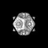
| Entry | Database: PDB / ID: 8co6 | ||||||||||||||||||
|---|---|---|---|---|---|---|---|---|---|---|---|---|---|---|---|---|---|---|---|
| Title | Subtomogram average of Immature Rotavirus TLP penton | ||||||||||||||||||
 Components Components |
| ||||||||||||||||||
 Keywords Keywords | VIRAL PROTEIN / Rotavirus / STA | ||||||||||||||||||
| Function / homology |  Function and homology information Function and homology informationviral intermediate capsid / host cell endoplasmic reticulum lumen / host cell rough endoplasmic reticulum / permeabilization of host organelle membrane involved in viral entry into host cell / T=13 icosahedral viral capsid / T=2 icosahedral viral capsid / host cytoskeleton / viral inner capsid / viral outer capsid / viral nucleocapsid ...viral intermediate capsid / host cell endoplasmic reticulum lumen / host cell rough endoplasmic reticulum / permeabilization of host organelle membrane involved in viral entry into host cell / T=13 icosahedral viral capsid / T=2 icosahedral viral capsid / host cytoskeleton / viral inner capsid / viral outer capsid / viral nucleocapsid / host cell endoplasmic reticulum-Golgi intermediate compartment / receptor-mediated virion attachment to host cell / host cell surface receptor binding / fusion of virus membrane with host plasma membrane / viral envelope / virion attachment to host cell / host cell plasma membrane / structural molecule activity / RNA binding / metal ion binding Similarity search - Function | ||||||||||||||||||
| Biological species |  Rotavirus A Rotavirus A | ||||||||||||||||||
| Method | ELECTRON MICROSCOPY / subtomogram averaging / cryo EM / Resolution: 4.7 Å | ||||||||||||||||||
 Authors Authors | Shah, P.N.M. / Stuart, D.I. | ||||||||||||||||||
| Funding support |  United Kingdom, 5items United Kingdom, 5items
| ||||||||||||||||||
 Citation Citation | Journal: Cell Host Microbe / Year: 2023 Title: Characterization of the rotavirus assembly pathway in situ using cryoelectron tomography. Authors: Pranav N M Shah / James B Gilchrist / Björn O Forsberg / Alister Burt / Andrew Howe / Shyamal Mosalaganti / William Wan / Julika Radecke / Yuriy Chaban / Geoff Sutton / David I Stuart / Mark Boyce /   Abstract: Rotavirus assembly is a complex process that involves the stepwise acquisition of protein layers in distinct intracellular locations to form the fully assembled particle. Understanding and ...Rotavirus assembly is a complex process that involves the stepwise acquisition of protein layers in distinct intracellular locations to form the fully assembled particle. Understanding and visualization of the assembly process has been hampered by the inaccessibility of unstable intermediates. We characterize the assembly pathway of group A rotaviruses observed in situ within cryo-preserved infected cells through the use of cryoelectron tomography of cellular lamellae. Our findings demonstrate that the viral polymerase VP1 recruits viral genomes during particle assembly, as revealed by infecting with a conditionally lethal mutant. Additionally, pharmacological inhibition to arrest the transiently enveloped stage uncovered a unique conformation of the VP4 spike. Subtomogram averaging provided atomic models of four intermediate states, including a pre-packaging single-layered intermediate, the double-layered particle, the transiently enveloped double-layered particle, and the fully assembled triple-layered virus particle. In summary, these complementary approaches enable us to elucidate the discrete steps involved in forming an intracellular rotavirus particle. | ||||||||||||||||||
| History |
|
- Structure visualization
| Structure viewer | Molecule: MolmilJmol/JSmol |
|---|
- Downloads & links
-Download
| PDBx/mmCIF format | 8co6.cif.gz | 1.9 MB | Display | PDBx/mmCIF format |
|---|---|---|---|---|
| PDB format | pdb8co6.ent.gz | Display | PDB format | |
| PDBx/mmJSON format | 8co6.json.gz | Tree view | PDBx/mmJSON format | |
| Others |  Other downloads Other downloads |
-Validation report
| Arichive directory | https://data.pdbj.org/pub/pdb/validation_reports/co/8co6ftp://data.pdbj.org/pub/pdb/validation_reports/co/8co6 | HTTPS FTP |
|---|
-Related structure data
| Related structure data |  16772MC  8bp8C  8coaC M: map data used to model this data C: citing same article ( |
|---|---|
| Similar structure data |


-Links
 PDBj
PDBj


- Assembly
| Deposited unit | 
|
|---|---|
| 1 | x 5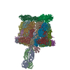
|
-Components
| #1: Protein | Mass: 86767.953 Da / Num. of mol.: 3 / Source method: isolated from a natural source / Source: (natural) Rotavirus A / References: UniProt: A0A1Q2TSK9#2: Protein | Mass: 37230.664 Da / Num. of mol.: 12 / Source method: isolated from a natural source / Source: (natural) Rotavirus A / References: UniProt: A0A1Q2TSM6#3: Protein | Mass: 44910.738 Da / Num. of mol.: 12 / Source method: isolated from a natural source / Source: (natural) Rotavirus A / References: UniProt: A2T3S6#4: Protein | Mass: 102412.961 Da / Num. of mol.: 2 / Source method: isolated from a natural source / Source: (natural) Rotavirus A / References: UniProt: A2T3R1Has protein modification | Y | |
|---|
-Experimental details
-Experiment
| Experiment | Method: ELECTRON MICROSCOPY |
|---|---|
| EM experiment | Aggregation state: PARTICLE / 3D reconstruction method: subtomogram averaging |
- Sample preparation
| Component | Name: Rotavirus A / Type: VIRUS / Entity ID: all / Source: NATURAL |
|---|---|
| Source (natural) | Organism: Rotavirus A |
| Details of virus | Empty: NO / Enveloped: NO / Isolate: STRAIN / Type: VIRION |
| Virus shell | Diameter: 1000 nm / Triangulation number (T number): 13 |
| Buffer solution | pH: 7.4 |
| Specimen | Embedding applied: NO / Shadowing applied: NO / Staining applied: NO / Vitrification applied: YES |
| Specimen support | Grid type: Quantifoil R2/2 |
| Vitrification | Cryogen name: ETHANE |
- Electron microscopy imaging
| Experimental equipment |  Model: Titan Krios / Image courtesy: FEI Company |
|---|---|
| Microscopy | Model: FEI TITAN KRIOS |
| Electron gun | Electron source:  FIELD EMISSION GUN / Accelerating voltage: 300 kV / Illumination mode: OTHER FIELD EMISSION GUN / Accelerating voltage: 300 kV / Illumination mode: OTHER |
| Electron lens | Mode: BRIGHT FIELD / Nominal defocus max: 3500 nm / Nominal defocus min: 1500 nm |
| Image recording | Electron dose: 2.2 e/Å2 / Avg electron dose per subtomogram: 90 e/Å2 / Film or detector model: FEI FALCON IV (4k x 4k) |
- Processing
| EM software |
| ||||||||||||||||||||||||
|---|---|---|---|---|---|---|---|---|---|---|---|---|---|---|---|---|---|---|---|---|---|---|---|---|---|
| CTF correction | Type: PHASE FLIPPING AND AMPLITUDE CORRECTION | ||||||||||||||||||||||||
| Symmetry | Point symmetry: C5 (5 fold cyclic) | ||||||||||||||||||||||||
| 3D reconstruction | Resolution: 4.7 Å / Resolution method: FSC 0.143 CUT-OFF / Num. of particles: 1961 / Symmetry type: POINT | ||||||||||||||||||||||||
| EM volume selection | Num. of tomograms: 85 / Num. of volumes extracted: 1961 | ||||||||||||||||||||||||
| Atomic model building | Protocol: FLEXIBLE FIT | ||||||||||||||||||||||||
| Atomic model building | Source name: PDB / Type: experimental model | ||||||||||||||||||||||||
| Refine LS restraints |
|
