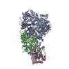
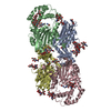
Journal: Science / Year: 2020 Title: In situ structural analysis of SARS-CoV-2 spike reveals flexibility mediated by three hinges. Authors: Beata Turoňová / Mateusz Sikora / Christoph Schürmann / Wim J H Hagen / Sonja Welsch / Florian E C Blanc / Sören von Bülow / Michael Gecht / Katrin Bagola / Cindy Hörner / Ger van ...Authors: Beata Turoňová / Mateusz Sikora / Christoph Schürmann / Wim J H Hagen / Sonja Welsch / Florian E C Blanc / Sören von Bülow / Michael Gecht / Katrin Bagola / Cindy Hörner / Ger van Zandbergen / Jonathan Landry / Nayara Trevisan Doimo de Azevedo / Shyamal Mosalaganti / Andre Schwarz / Roberto Covino / Michael D Mühlebach / Gerhard Hummer / Jacomine Krijnse Locker / Martin Beck / Abstract: The spike protein (S) of severe acute respiratory syndrome coronavirus 2 (SARS-CoV-2) is required for cell entry and is the primary focus for vaccine development. In this study, we combined cryo- ...The spike protein (S) of severe acute respiratory syndrome coronavirus 2 (SARS-CoV-2) is required for cell entry and is the primary focus for vaccine development. In this study, we combined cryo-electron tomography, subtomogram averaging, and molecular dynamics simulations to structurally analyze S in situ. Compared with the recombinant S, the viral S was more heavily glycosylated and occurred mostly in the closed prefusion conformation. We show that the stalk domain of S contains three hinges, giving the head unexpected orientational freedom. We propose that the hinges allow S to scan the host cell surface, shielded from antibodies by an extensive glycan coat. The structure of native S contributes to our understanding of SARS-CoV-2 infection and potentially to the development of safe vaccines.
History
Deposition
Jun 24, 2020
-
Header (metadata) release
Mar 17, 2021
-
Map release
Mar 17, 2021
-
Update
Dec 21, 2022
-
Current status
Dec 21, 2022
Processing site: PDBe / Status: Released
-
Structure visualization
Movie
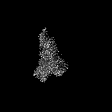
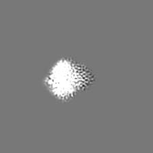
Surface view with section colored by density value
EMPIAR-10453 (Title: Cryo electron tomography - tilt-series of Sars-Cov-2 / Data size: 469.8 / Data #1: Unaligned tilt-series of Sars-Cov-2 [tilt series])
In the structure databanks used in Yorodumi, some data are registered as the other names, "COVID-19 virus" and "2019-nCoV". Here are the details of the virus and the list of structure data.
Jan 31, 2019. EMDB accession codes are about to change! (news from PDBe EMDB page)
EMDB accession codes are about to change! (news from PDBe EMDB page)
The allocation of 4 digits for EMDB accession codes will soon come to an end. Whilst these codes will remain in use, new EMDB accession codes will include an additional digit and will expand incrementally as the available range of codes is exhausted. The current 4-digit format prefixed with “EMD-” (i.e. EMD-XXXX) will advance to a 5-digit format (i.e. EMD-XXXXX), and so on. It is currently estimated that the 4-digit codes will be depleted around Spring 2019, at which point the 5-digit format will come into force.
The EM Navigator/Yorodumi systems omit the EMD- prefix.
Related info.:Q: What is EMD? / ID/Accession-code notation in Yorodumi/EM Navigator
Yorodumi is a browser for structure data from EMDB, PDB, SASBDB, etc.
This page is also the successor to EM Navigator detail page, and also detail information page/front-end page for Omokage search.
The word "yorodu" (or yorozu) is an old Japanese word meaning "ten thousand". "mi" (miru) is to see.
Related info.:EMDB / PDB / SASBDB / Comparison of 3 databanks / Yorodumi Search / Aug 31, 2016. New EM Navigator & Yorodumi / Yorodumi Papers / Jmol/JSmol / Function and homology information / Changes in new EM Navigator and Yorodumi
 Movie
Movie Controller

 Yorodumi
 Open data
 Basic information
 Map data
 Sample
 Function and homology information
 Severe acute respiratory syndrome coronavirus 2
 Authors
 Citation
 Structure visualization
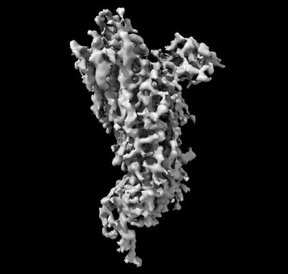
 Downloads & links
 emd_11223.png
 http://ftp.pdbj.org/pub/emdb/structures/EMD-11223


 Z (Sec.)
 Y (Row.)
 X (Col.)


 Sample components
 Processing
 Electron microscopy
 FIELD EMISSION GUN