 Movie
Movie Controller


+ Open data

- Basic information
| Entry | Database: PDB / ID: 3jca | ||||||
|---|---|---|---|---|---|---|---|
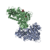
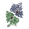
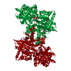
| Title | Core model of the Mouse Mammary Tumor Virus intasome | ||||||
 Components Components |
| ||||||
 Keywords Keywords | VIRAL PROTEIN / integration / retrovirus / integrase / intasome | ||||||
| Function / homology |  Function and homology information Function and homology informationdUTP diphosphatase / dUTP diphosphatase activity / Hydrolases; Acting on peptide bonds (peptidases); Aspartic endopeptidases / ribonuclease H / DNA integration / viral genome integration into host DNA / establishment of integrated proviral latency / RNA-directed DNA polymerase / RNA stem-loop binding / RNA-directed DNA polymerase activity ...dUTP diphosphatase / dUTP diphosphatase activity / Hydrolases; Acting on peptide bonds (peptidases); Aspartic endopeptidases / ribonuclease H / DNA integration / viral genome integration into host DNA / establishment of integrated proviral latency / RNA-directed DNA polymerase / RNA stem-loop binding / RNA-directed DNA polymerase activity / RNA-DNA hybrid ribonuclease activity / Transferases; Transferring phosphorus-containing groups; Nucleotidyltransferases / viral nucleocapsid / DNA recombination / DNA-directed DNA polymerase / structural constituent of virion / aspartic-type endopeptidase activity / Hydrolases; Acting on ester bonds / DNA-directed DNA polymerase activity / viral translational frameshifting / symbiont entry into host cell / proteolysis / DNA binding / zinc ion binding Similarity search - Function | ||||||
| Biological species |  Mouse mammary tumor virus Mouse mammary tumor virus | ||||||
| Method | ELECTRON MICROSCOPY / single particle reconstruction / cryo EM / Resolution: 4.8 Å | ||||||
 Authors Authors | Lyumkis, D.L. / Ballandras-Colas, A. / Brown, M. / Cook, N.J. / Dewdney, T.G. / Demeler, B. / Cherepanov, P. / Engelman, A.N. | ||||||
 Citation Citation | Journal: Nature / Year: 2016 Title: Cryo-EM reveals a novel octameric integrase structure for betaretroviral intasome function. Authors: Allison Ballandras-Colas / Monica Brown / Nicola J Cook / Tamaria G Dewdney / Borries Demeler / Peter Cherepanov / Dmitry Lyumkis / Alan N Engelman /   Abstract: Retroviral integrase catalyses the integration of viral DNA into host target DNA, which is an essential step in the life cycle of all retroviruses. Previous structural characterization of integrase- ...Retroviral integrase catalyses the integration of viral DNA into host target DNA, which is an essential step in the life cycle of all retroviruses. Previous structural characterization of integrase-viral DNA complexes, or intasomes, from the spumavirus prototype foamy virus revealed a functional integrase tetramer, and it is generally believed that intasomes derived from other retroviral genera use tetrameric integrase. However, the intasomes of orthoretroviruses, which include all known pathogenic species, have not been characterized structurally. Here, using single-particle cryo-electron microscopy and X-ray crystallography, we determine an unexpected octameric integrase architecture for the intasome of the betaretrovirus mouse mammary tumour virus. The structure is composed of two core integrase dimers, which interact with the viral DNA ends and structurally mimic the integrase tetramer of prototype foamy virus, and two flanking integrase dimers that engage the core structure via their integrase carboxy-terminal domains. Contrary to the belief that tetrameric integrase components are sufficient to catalyse integration, the flanking integrase dimers were necessary for mouse mammary tumour virus integrase activity. The integrase octamer solves a conundrum for betaretroviruses as well as alpharetroviruses by providing critical carboxy-terminal domains to the intasome core that cannot be provided in cis because of evolutionarily restrictive catalytic core domain-carboxy-terminal domain linker regions. The octameric architecture of the intasome of mouse mammary tumour virus provides new insight into the structural basis of retroviral DNA integration. | ||||||
| History |
|
- Structure visualization
| Movie |
Movie viewer |
|---|---|
| Structure viewer | Molecule: MolmilJmol/JSmol |
- Downloads & links
-Download
| PDBx/mmCIF format | 3jca.cif.gz | 292.4 KB | Display | PDBx/mmCIF format |
|---|---|---|---|---|
| PDB format | pdb3jca.ent.gz | 232.7 KB | Display | PDB format |
| PDBx/mmJSON format | 3jca.json.gz | Tree view | PDBx/mmJSON format | |
| Others |  Other downloads Other downloads |
-Validation report
| Arichive directory | https://data.pdbj.org/pub/pdb/validation_reports/jc/3jcaftp://data.pdbj.org/pub/pdb/validation_reports/jc/3jca | HTTPS FTP |
|---|
-Related structure data
| Related structure data | 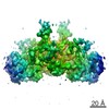 6441MC 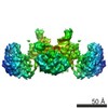 6440C  5cz1C  5cz2C  5d7uC M: map data used to model this data C: citing same article ( |
|---|---|
| Similar structure data |


-Links
 PDBj
PDBj


- Assembly
| Deposited unit | 
|
|---|---|
| 1 |
|
-Components
| #1: Protein | Mass: 30034.287 Da / Num. of mol.: 4 / Fragment: UNP residues 1437-1701 Source method: isolated from a genetically manipulated source Source: (gene. exp.) Mouse mammary tumor virus / Plasmid: pET15b / Production host:  #2: Protein/peptide | Mass: 5571.474 Da / Num. of mol.: 4 / Fragment: C-terminal domain (UNP residues 1653-1701) Source method: isolated from a genetically manipulated source Source: (gene. exp.) Mouse mammary tumor virus / Plasmid: pET15b / Production host: #3: DNA chain | Mass: 6738.343 Da / Num. of mol.: 2 / Source method: obtained synthetically / Details: pre-cleaved viral DNA 3' strand / Source: (synth.) Mouse mammary tumor virus#4: DNA chain | Mass: 6160.968 Da / Num. of mol.: 2 / Source method: obtained synthetically / Details: pre-cleaved viral DNA 5' strand / Source: (synth.) Mouse mammary tumor virus#5: Chemical | ChemComp-ZN /   Mass: 65.409 Da / Num. of mol.: 4 / Source method: obtained synthetically / Formula: Zn Mass: 65.409 Da / Num. of mol.: 4 / Source method: obtained synthetically / Formula: Zn |
|---|
-Experimental details
-Experiment
| Experiment | Method: ELECTRON MICROSCOPY |
|---|---|
| EM experiment | Aggregation state: PARTICLE / 3D reconstruction method: single particle reconstruction |
- Sample preparation
| Component |
| ||||||||||||||||||||
|---|---|---|---|---|---|---|---|---|---|---|---|---|---|---|---|---|---|---|---|---|---|
| Molecular weight | Value: 2 MDa / Experimental value: NO | ||||||||||||||||||||
| Buffer solution | Name: size-exclusion buffer / pH: 7.4 / Details: size-exclusion buffer | ||||||||||||||||||||
| Specimen | Embedding applied: NO / Shadowing applied: NO / Staining applied: NO / Vitrification applied: YES | ||||||||||||||||||||
| Specimen support | Details: 400 mesh C-flat 1.2/1.3-4C, plasma-treated for 6 seconds | ||||||||||||||||||||
| Vitrification | Instrument: HOMEMADE PLUNGER / Cryogen name: ETHANE / Temp: 77 K / Humidity: 90 % Details: Sample containing MMTV intasomes in SEC buffer (to which the detergent NP-40 was added to a final concentration of 0.05%) was applied onto a freshly plasma-treated (6s, Gatan Solarus plasma ...Details: Sample containing MMTV intasomes in SEC buffer (to which the detergent NP-40 was added to a final concentration of 0.05%) was applied onto a freshly plasma-treated (6s, Gatan Solarus plasma cleaner) holey carbon C-flat grid (CF-1.2/1.3-4C, Protochips, Inc.), allowing the sample to adsorb for 30 seconds and then plunge-freezing in liquid ethane using a manual cryo-plunger in an ambient environment of 4 degrees C. Method: 3 uL sample was applied to grid, adsorbed for 30 seconds, blotted, and plunge-frozen in liquid ethane. |
- Electron microscopy imaging
| Experimental equipment |  Model: Titan Krios / Image courtesy: FEI Company |
|---|---|
| Microscopy | Model: FEI TITAN KRIOS / Date: Apr 15, 2015 |
| Electron gun | Electron source:  FIELD EMISSION GUN / Accelerating voltage: 300 kV / Illumination mode: FLOOD BEAM FIELD EMISSION GUN / Accelerating voltage: 300 kV / Illumination mode: FLOOD BEAM |
| Electron lens | Mode: BRIGHT FIELD / Nominal magnification: 22500 X / Calibrated magnification: 38167 X / Nominal defocus max: 4000 nm / Nominal defocus min: 1000 nm / Cs: 2.7 mm Astigmatism: Objective lens astigmatism was corrected using Leginon, and coma-free alignment was established. |
| Specimen holder | Specimen holder model: FEI TITAN KRIOS AUTOGRID HOLDER |
| Image recording | Electron dose: 40 e/Å2 / Film or detector model: GATAN K2 (4k x 4k) |
| Image scans | Num. digital images: 2714 |
- Processing
| EM software |
| ||||||||||||||||||||||||||||
|---|---|---|---|---|---|---|---|---|---|---|---|---|---|---|---|---|---|---|---|---|---|---|---|---|---|---|---|---|---|
| CTF correction | Details: each particle | ||||||||||||||||||||||||||||
| Symmetry | Point symmetry: C2 (2 fold cyclic) | ||||||||||||||||||||||||||||
| 3D reconstruction | Method: projection matching / Resolution: 4.8 Å / Resolution method: FSC 0.143 CUT-OFF / Num. of particles: 30307 / Nominal pixel size: 1.31 Å / Actual pixel size: 1.31 Å Details: Local FSC values range from 4-5 A. (Single particle details: IN-NTD and IN-CCD domains of flanking INs 5-8 were computationally removed using Relion after assignment of Euler angles to full ...Details: Local FSC values range from 4-5 A. (Single particle details: IN-NTD and IN-CCD domains of flanking INs 5-8 were computationally removed using Relion after assignment of Euler angles to full octameric particles and masking out of flanking regions.) (Single particle - Applied symmetry: C2) Symmetry type: POINT | ||||||||||||||||||||||||||||
| Atomic model building | B value: 300 / Protocol: OTHER / Space: REAL / Target criteria: FSC, Rosetta energy Details: REFINEMENT PROTOCOL--real-space refinement DETAILS--Initial components solved by X-ray were rigid-body docked. Linker regions were built de novo from the EM density. The full model was ...Details: REFINEMENT PROTOCOL--real-space refinement DETAILS--Initial components solved by X-ray were rigid-body docked. Linker regions were built de novo from the EM density. The full model was refined using real-space methods in Rosetta. | ||||||||||||||||||||||||||||
| Atomic model building |
| ||||||||||||||||||||||||||||
| Refinement step | Cycle: LAST
|
