 Movie
Movie Controller


+ Open data

- Basic information
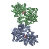
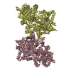
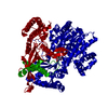
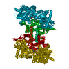
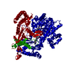
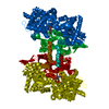
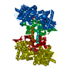
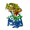
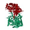
| Entry | Database: PDB / ID: 1fa9 | ||||||
|---|---|---|---|---|---|---|---|
| Title | HUMAN LIVER GLYCOGEN PHOSPHORYLASE A COMPLEXED WITH AMP | ||||||
 Components Components | GLYCOGEN PHOSPHORYLASE, LIVER FORM | ||||||
 Keywords Keywords | TRANSFERASE / protein-ligand complex / allosteric protein / phosphorylated protein | ||||||
| Function / homology |  Function and homology information Function and homology informationpurine nucleobase binding / vitamin binding / D-glucose binding / bile acid binding / glycogen phosphorylase / glycogen phosphorylase activity / glycogen catabolic process / glycogen metabolic process / AMP binding / Glycogen breakdown (glycogenolysis) ...purine nucleobase binding / vitamin binding / D-glucose binding / bile acid binding / glycogen phosphorylase / glycogen phosphorylase activity / glycogen catabolic process / glycogen metabolic process / AMP binding / Glycogen breakdown (glycogenolysis) / pyridoxal phosphate binding / glucose homeostasis / secretory granule lumen / ficolin-1-rich granule lumen / Neutrophil degranulation / extracellular exosome / extracellular region / ATP binding / identical protein binding / cytosol / cytoplasm Similarity search - Function | ||||||
| Biological species |  Homo sapiens (human) Homo sapiens (human) | ||||||
| Method |  X-RAY DIFFRACTION / MOLECULAR REPLACEMENT / Resolution: 2.4 Å X-RAY DIFFRACTION / MOLECULAR REPLACEMENT / Resolution: 2.4 Å | ||||||
 Authors Authors | Rath, V.L. / Ammirati, M. / LeMotte, P.K. / Fennell, K.F. / Mansour, M.N. / Danley, D.E. / Hynes, T.R. / Schulte, G.K. / Wasilko, D.J. / Pandit, J. | ||||||
 Citation Citation | Journal: Mol.Cell / Year: 2000 Title: Activation of human liver glycogen phosphorylase by alteration of the secondary structure and packing of the catalytic core. Authors: Rath, V.L. / Ammirati, M. / LeMotte, P.K. / Fennell, K.F. / Mansour, M.N. / Danley, D.E. / Hynes, T.R. / Schulte, G.K. / Wasilko, D.J. / Pandit, J. | ||||||
| History |
|
- Structure visualization
| Structure viewer | Molecule: MolmilJmol/JSmol |
|---|
- Downloads & links
-Download
| PDBx/mmCIF format | 1fa9.cif.gz | 187.8 KB | Display | PDBx/mmCIF format |
|---|---|---|---|---|
| PDB format | pdb1fa9.ent.gz | 146.8 KB | Display | PDB format |
| PDBx/mmJSON format | 1fa9.json.gz | Tree view | PDBx/mmJSON format | |
| Others |  Other downloads Other downloads |
-Validation report
| Arichive directory | https://data.pdbj.org/pub/pdb/validation_reports/fa/1fa9ftp://data.pdbj.org/pub/pdb/validation_reports/fa/1fa9 | HTTPS FTP |
|---|
-Related structure data
| Related structure data | 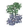 1fc0C  1gpaS C: citing same article ( S: Starting model for refinement |
|---|---|
| Similar structure data |
-Links
 PDBj
PDBj


- Assembly
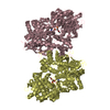
| Deposited unit | 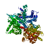
| ||||||||
|---|---|---|---|---|---|---|---|---|---|
| 1 | 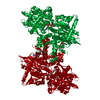
| ||||||||
| Unit cell |
| ||||||||
| Details | The biological assembly is a dimer constructed from chain A a symmetry partner generated by the two-fold. |
-Components
| #1: Protein | Mass: 97225.250 Da / Num. of mol.: 1 Source method: isolated from a genetically manipulated source Source: (gene. exp.) Homo sapiens (human) / Tissue: LIVER / Plasmid: PKK2332 / Production host:  |
|---|---|
| #2: Sugar | ChemComp-GLC /   Type: D-saccharide, alpha linking / Mass: 180.156 Da / Num. of mol.: 1 Type: D-saccharide, alpha linking / Mass: 180.156 Da / Num. of mol.: 1Source method: isolated from a genetically manipulated source Formula: C6H12O6 |
| #3: Chemical | ChemComp-AMP /   Mass: 347.221 Da / Num. of mol.: 1 / Source method: obtained synthetically / Formula: C10H14N5O7P / Comment: AMP*YM Mass: 347.221 Da / Num. of mol.: 1 / Source method: obtained synthetically / Formula: C10H14N5O7P / Comment: AMP*YM |
| #4: Chemical | ChemComp-PLP /   Mass: 247.142 Da / Num. of mol.: 1 / Source method: obtained synthetically / Formula: C8H10NO6P Mass: 247.142 Da / Num. of mol.: 1 / Source method: obtained synthetically / Formula: C8H10NO6P |
| #5: Water | ChemComp-HOH /  Mass: 18.015 Da / Num. of mol.: 242 / Source method: isolated from a natural source / Formula: H2O Mass: 18.015 Da / Num. of mol.: 242 / Source method: isolated from a natural source / Formula: H2O |
-Experimental details
-Experiment
| Experiment | Method: X-RAY DIFFRACTION / Number of used crystals: 1 |
|---|
- Sample preparation
| Crystal | Density Matthews: 2.91 Å3/Da / Density % sol: 57.72 % | ||||||||||||||||||||||||||||||||||||||||
|---|---|---|---|---|---|---|---|---|---|---|---|---|---|---|---|---|---|---|---|---|---|---|---|---|---|---|---|---|---|---|---|---|---|---|---|---|---|---|---|---|---|
| Crystal grow | Temperature: 298 K / Method: vapor diffusion, hanging drop / pH: 8.5 Details: Peg 8000, Tris/HCl, AMP, glucose, pH 8.5, VAPOR DIFFUSION, HANGING DROP, temperature 25K | ||||||||||||||||||||||||||||||||||||||||
| Crystal grow | *PLUS pH: 6.8 | ||||||||||||||||||||||||||||||||||||||||
| Components of the solutions | *PLUS
|
-Data collection
| Diffraction | Mean temperature: 100 K |
|---|---|
| Diffraction source | Source: ROTATING ANODE / Type: RIGAKU RU200 / Wavelength: 1.5418 |
| Detector | Type: RIGAKU RAXIS IIC / Detector: IMAGE PLATE / Date: Jul 19, 1994 |
| Radiation | Protocol: SINGLE WAVELENGTH / Monochromatic (M) / Laue (L): M / Scattering type: x-ray |
| Radiation wavelength | Wavelength: 1.5418 Å / Relative weight: 1 |
| Reflection | Resolution: 2.4→50 Å / Num. all: 44772 / Num. obs: 43732 / % possible obs: 98.5 % / Observed criterion σ(F): 0 / Observed criterion σ(I): -2 / Redundancy: 9 % / Biso Wilson estimate: 38.3 Å2 / Rmerge(I) obs: 0.057 / Net I/σ(I): 22.9 |
| Reflection shell | Resolution: 2.4→2.49 Å / Redundancy: 2 % / Rmerge(I) obs: 0.495 / % possible all: 91.8 |
| Reflection | *PLUS Num. obs: 53400 |
| Reflection shell | *PLUS % possible obs: 91.8 % |
- Processing
| Software |
| |||||||||||||||||||||||||
|---|---|---|---|---|---|---|---|---|---|---|---|---|---|---|---|---|---|---|---|---|---|---|---|---|---|---|
| Refinement | Method to determine structure: MOLECULAR REPLACEMENT Starting model: 1GPA Resolution: 2.4→50 Å / Cross valid method: X-PLOR / σ(F): 0 / σ(I): 0 / Stereochemistry target values: Engh & Huber
| |||||||||||||||||||||||||
| Refinement step | Cycle: LAST / Resolution: 2.4→50 Å
| |||||||||||||||||||||||||
| Refine LS restraints |
| |||||||||||||||||||||||||
| LS refinement shell | Resolution: 2.4→2.42 Å / Rfactor Rfree: 0.476 / Rfactor Rwork: 0.353 | |||||||||||||||||||||||||
| Software | *PLUS Name: X-PLOR / Version: 3.1 / Classification: refinement | |||||||||||||||||||||||||
| Refinement | *PLUS Highest resolution: 2.4 Å / Lowest resolution: 50 Å / σ(F): 0 / % reflection Rfree: 10 % | |||||||||||||||||||||||||
| Solvent computation | *PLUS | |||||||||||||||||||||||||
| Displacement parameters | *PLUS | |||||||||||||||||||||||||
| Refine LS restraints | *PLUS
| |||||||||||||||||||||||||
| LS refinement shell | *PLUS Highest resolution: 2.4 Å / Rfactor Rfree: 0.476 / Rfactor Rwork: 0.353 / Rfactor obs: 0.353 |
