 Movie
Movie Controller

[English] 日本語
 Yorodumi
- PDB-1gpy: CRYSTALLOGRAPHIC BINDING STUDIES ON THE ALLOSTERIC INHIBITOR GLUC... -
+ Open data

- Basic information
| Entry | Database: PDB / ID: 1gpy | ||||||
|---|---|---|---|---|---|---|---|
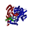
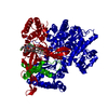
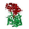
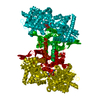
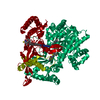
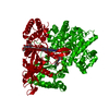
| Title | CRYSTALLOGRAPHIC BINDING STUDIES ON THE ALLOSTERIC INHIBITOR GLUCOSE-6-PHOSPHATE TO T STATE GLYCOGEN PHOSPHORYLASE B | ||||||
 Components Components | GLYCOGEN PHOSPHORYLASE B | ||||||
 Keywords Keywords | GLYCOGEN PHOSPHORYLASE | ||||||
| Function / homology |  Function and homology information Function and homology informationglycogen phosphorylase / glycogen phosphorylase activity / glycogen catabolic process / skeletal muscle myofibril / pyridoxal phosphate binding / nucleotide binding Similarity search - Function | ||||||
| Biological species |  | ||||||
| Method |  X-RAY DIFFRACTION / Resolution: 2.4 Å X-RAY DIFFRACTION / Resolution: 2.4 Å | ||||||
 Authors Authors | Johnson, L.N. | ||||||
 Citation Citation | Journal: J.Mol.Biol. / Year: 1993 Title: Crystallographic binding studies on the allosteric inhibitor glucose-6-phosphate to T state glycogen phosphorylase b. Authors: Johnson, L.N. / Snape, P. / Martin, J.L. / Acharya, K.R. / Barford, D. / Oikonomakos, N.G. #1: Journal: Glycogen Phosphorylase B: Description of the Protein StructureYear: 1991 Title: Glycogen Phosphorylase B: Description of the Protein Structure Authors: Acharya, K.R. / Stuart, D.I. / Varvill, K.M. / Johnson, L.N. #2: Journal: J.Mol.Biol. / Year: 1991Title: Structural Mechanism for Glycogen Phosphorylase Control by Phosphorylation and AMP Authors: Barford, D. / Hu, S.-H. / Johnson, L.N. | ||||||
| History |
|
- Structure visualization
| Structure viewer | Molecule: MolmilJmol/JSmol |
|---|
- Downloads & links
-Download
| PDBx/mmCIF format | 1gpy.cif.gz | 199.3 KB | Display | PDBx/mmCIF format |
|---|---|---|---|---|
| PDB format | pdb1gpy.ent.gz | 155.1 KB | Display | PDB format |
| PDBx/mmJSON format | 1gpy.json.gz | Tree view | PDBx/mmJSON format | |
| Others |  Other downloads Other downloads |
-Validation report
| Arichive directory | https://data.pdbj.org/pub/pdb/validation_reports/gp/1gpyftp://data.pdbj.org/pub/pdb/validation_reports/gp/1gpy | HTTPS FTP |
|---|
-Related structure data
| Similar structure data |
|---|


-Links
 PDBj
PDBj
- Assembly
| Deposited unit | 
| ||||||||
|---|---|---|---|---|---|---|---|---|---|
| 1 | 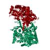
| ||||||||
| Unit cell |
| ||||||||
| Atom site foot note | 1: RESIDUE 837 IS A CIS PROLINE. |
-Components
| #1: Protein | Mass: 97291.203 Da / Num. of mol.: 1 Source method: isolated from a genetically manipulated source Source: (gene. exp.) |
|---|---|
| #2: Sugar | ChemComp-G6P /   Type: D-saccharide, alpha linking / Mass: 260.136 Da / Num. of mol.: 1 Type: D-saccharide, alpha linking / Mass: 260.136 Da / Num. of mol.: 1Source method: isolated from a genetically manipulated source Formula: C6H13O9P |
| #3: Chemical | ChemComp-PLP /   Mass: 247.142 Da / Num. of mol.: 1 / Source method: obtained synthetically / Formula: C8H10NO6P Mass: 247.142 Da / Num. of mol.: 1 / Source method: obtained synthetically / Formula: C8H10NO6P |
| #4: Water | ChemComp-HOH /  Mass: 18.015 Da / Num. of mol.: 673 / Source method: isolated from a natural source / Formula: H2O Mass: 18.015 Da / Num. of mol.: 673 / Source method: isolated from a natural source / Formula: H2O |
| Has protein modification | N |
| Nonpolymer details | PLP FORMS A SCHIFF'S BASE WITH NZ OF LYS 680. |
-Experimental details
-Experiment
| Experiment | Method: X-RAY DIFFRACTION |
|---|
- Sample preparation
| Crystal | Density Matthews: 2.47 Å3/Da / Density % sol: 50.13 % | ||||||||||||||||||||||||||||||||||||||||||
|---|---|---|---|---|---|---|---|---|---|---|---|---|---|---|---|---|---|---|---|---|---|---|---|---|---|---|---|---|---|---|---|---|---|---|---|---|---|---|---|---|---|---|---|
| Crystal grow | *PLUS pH: 6.7 / Method: unknown | ||||||||||||||||||||||||||||||||||||||||||
| Components of the solutions | *PLUS
|
-Data collection
| Radiation | Scattering type: x-ray |
|---|---|
| Radiation wavelength | Relative weight: 1 |
| Reflection | *PLUS Highest resolution: 2.3 Å / Num. obs: 35718 / % possible obs: 78.6 % / Num. measured all: 90321 / Rmerge(I) obs: 0.073 |
- Processing
| Software |
| ||||||||||||||||||||||||||||||||||||||||||||||||||||||||||||
|---|---|---|---|---|---|---|---|---|---|---|---|---|---|---|---|---|---|---|---|---|---|---|---|---|---|---|---|---|---|---|---|---|---|---|---|---|---|---|---|---|---|---|---|---|---|---|---|---|---|---|---|---|---|---|---|---|---|---|---|---|---|
| Refinement | Rfactor Rwork: 0.203 / Rfactor obs: 0.203 / Highest resolution: 2.4 Å | ||||||||||||||||||||||||||||||||||||||||||||||||||||||||||||
| Refinement step | Cycle: LAST / Highest resolution: 2.4 Å
| ||||||||||||||||||||||||||||||||||||||||||||||||||||||||||||
| Refine LS restraints |
| ||||||||||||||||||||||||||||||||||||||||||||||||||||||||||||
| Refinement | *PLUS Highest resolution: 2.3 Å / Lowest resolution: 8 Å / Num. reflection all: 32624 / σ(F): 0 / Rfactor all: 0.203 | ||||||||||||||||||||||||||||||||||||||||||||||||||||||||||||
| Solvent computation | *PLUS | ||||||||||||||||||||||||||||||||||||||||||||||||||||||||||||
| Displacement parameters | *PLUS | ||||||||||||||||||||||||||||||||||||||||||||||||||||||||||||
| Refine LS restraints | *PLUS Type: x_angle_d / Dev ideal: 4 |
