 Movie
Movie Controller

[English] 日本語
 Yorodumi
- PDB-1k08: Crystallographic Binding Study of 10 mM N-benzoyl-N'-beta-D-gluco... -
+ Open data

- Basic information
| Entry | Database: PDB / ID: 1k08 | ||||||
|---|---|---|---|---|---|---|---|
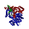
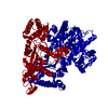
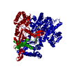
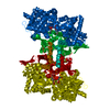
| Title | Crystallographic Binding Study of 10 mM N-benzoyl-N'-beta-D-glucopyranosyl urea to glycogen phosphorylase b | ||||||
 Components Components | Glycogen Phosphorylase | ||||||
 Keywords Keywords | TRANSFERASE / glycogen phosphorylase / catalytic site / new allosteric site | ||||||
| Function / homology |  Function and homology information Function and homology informationglycogen phosphorylase / glycogen phosphorylase activity / glycogen catabolic process / skeletal muscle myofibril / pyridoxal phosphate binding / nucleotide binding Similarity search - Function | ||||||
| Biological species |  | ||||||
| Method |  X-RAY DIFFRACTION / FOURIER SYNTHESIS / Resolution: 2.26 Å X-RAY DIFFRACTION / FOURIER SYNTHESIS / Resolution: 2.26 Å | ||||||
 Authors Authors | Oikonomakos, N.G. / Kosmopoulou, M. / Zographos, S.E. / Leonidas, D.D. / Chrysina, E.D. / Somsak, L. / Nagy, V. / Praly, J.P. / Docsa, T. / Toth, B. / Gergely, P. | ||||||
 Citation Citation | Journal: Eur.J.Biochem. / Year: 2002 Title: Binding of N-acetyl-N '-beta-D-glucopyranosyl urea and N-benzoyl-N '-beta-D-glucopyranosyl urea to glycogen phosphorylase b: kinetic and crystallographic studies. Authors: Oikonomakos, N.G. / Kosmopoulou, M. / Zographos, S.E. / Leonidas, D.D. / Chrysina, E.D. / Somsak, L. / Nagy, V. / Praly, J.P. / Docsa, T. / Toth, B. / Gergely, P. #1: Journal: Structure / Year: 2000Title: A New Allosteric Site in Glycogen Phosphorylase B as a Target for Drug Interactions Authors: Oikonomakos, N.G. / Skamnaki, V.T. / Tsitsanou, K.E. / Gavalas, N.G. / Johnson, L.N. | ||||||
| History |
|
- Structure visualization
| Structure viewer | Molecule: MolmilJmol/JSmol |
|---|
- Downloads & links
-Download
| PDBx/mmCIF format | 1k08.cif.gz | 182.2 KB | Display | PDBx/mmCIF format |
|---|---|---|---|---|
| PDB format | pdb1k08.ent.gz | 143.2 KB | Display | PDB format |
| PDBx/mmJSON format | 1k08.json.gz | Tree view | PDBx/mmJSON format | |
| Others |  Other downloads Other downloads |
-Validation report
| Arichive directory | https://data.pdbj.org/pub/pdb/validation_reports/k0/1k08ftp://data.pdbj.org/pub/pdb/validation_reports/k0/1k08 | HTTPS FTP |
|---|
-Related structure data
| Related structure data |  1k06C  1ktiC  1hlfS C: citing same article ( S: Starting model for refinement |
|---|---|
| Similar structure data |


-Links
 PDBj
PDBj
- Assembly
| Deposited unit | 
| ||||||||
|---|---|---|---|---|---|---|---|---|---|
| 1 | 
| ||||||||
| Unit cell |
| ||||||||
| Components on special symmetry positions |
|
-Components
| #1: Protein | Mass: 97291.203 Da / Num. of mol.: 1 / Source method: isolated from a natural source / Source: (natural) | ||||
|---|---|---|---|---|---|
| #2: Sugar |   Type: D-saccharide / Mass: 326.302 Da / Num. of mol.: 2 / Source method: obtained synthetically / Formula: C14H18N2O7 Type: D-saccharide / Mass: 326.302 Da / Num. of mol.: 2 / Source method: obtained synthetically / Formula: C14H18N2O7#3: Chemical | ChemComp-PLP / |   Mass: 247.142 Da / Num. of mol.: 1 / Source method: obtained synthetically / Formula: C8H10NO6P Mass: 247.142 Da / Num. of mol.: 1 / Source method: obtained synthetically / Formula: C8H10NO6P#4: Water | ChemComp-HOH / |  Mass: 18.015 Da / Num. of mol.: 249 / Source method: isolated from a natural source / Formula: H2O Mass: 18.015 Da / Num. of mol.: 249 / Source method: isolated from a natural source / Formula: H2O |
-Experimental details
-Experiment
| Experiment | Method: X-RAY DIFFRACTION / Number of used crystals: 1 |
|---|
- Sample preparation
| Crystal | Density Matthews: 2.47 Å3/Da / Density % sol: 50.23 % |
|---|---|
| Crystal grow | Temperature: 287 K / Method: small tubes / pH: 6.7 Details: BES, EDTA, 10mM N-benzoyl-N'-beta-D-glucopyranosyl urea, pH 6.7, SMALL TUBES, temperature 287K |
| Crystal grow | *PLUS Details: Oikonomakos, N.G., (1985) Biochim. Biophys. Acta, 832, 248. |
-Data collection
| Diffraction | Mean temperature: 298 K |
|---|---|
| Diffraction source | Source: ROTATING ANODE / Type: RIGAKU / Wavelength: 1.5418 Å |
| Detector | Type: RIGAKU RAXIS IV / Detector: IMAGE PLATE / Date: Apr 2, 2001 / Details: mirrors |
| Radiation | Monochromator: Yale mirrors / Protocol: SINGLE WAVELENGTH / Monochromatic (M) / Laue (L): M / Scattering type: x-ray |
| Radiation wavelength | Wavelength: 1.5418 Å / Relative weight: 1 |
| Reflection | Resolution: 2.26→28.75 Å / Num. all: 44261 / Num. obs: 44361 / % possible obs: 95.8 % / Observed criterion σ(F): 0 / Observed criterion σ(I): -3 / Redundancy: 5.3 % / Biso Wilson estimate: 28 Å2 / Rsym value: 9.6 / Net I/σ(I): 10.9 |
| Reflection shell | Resolution: 2.26→2.3 Å / Redundancy: 4.2 % / Mean I/σ(I) obs: 2.6 / Num. unique all: 1976 / Rsym value: 42.4 / % possible all: 86.5 |
| Reflection | *PLUS Num. measured all: 391254 / Rmerge(I) obs: 0.096 |
| Reflection shell | *PLUS % possible obs: 86.5 % / Rmerge(I) obs: 0.424 |
- Processing
| Software |
| ||||||||||||||||||||||||||||||||||||||||||||||||||||||||||||||||||||||||||||||||
|---|---|---|---|---|---|---|---|---|---|---|---|---|---|---|---|---|---|---|---|---|---|---|---|---|---|---|---|---|---|---|---|---|---|---|---|---|---|---|---|---|---|---|---|---|---|---|---|---|---|---|---|---|---|---|---|---|---|---|---|---|---|---|---|---|---|---|---|---|---|---|---|---|---|---|---|---|---|---|---|---|---|
| Refinement | Method to determine structure: FOURIER SYNTHESIS Starting model: PDB ENTRY 1HLF Resolution: 2.26→28.75 Å / Rfactor Rfree error: 0.004 / Data cutoff high absF: 10000000 / Data cutoff low absF: 0.001 / Isotropic thermal model: RESTRAINED / Cross valid method: THROUGHOUT / σ(F): 0 / Stereochemistry target values: Engh & Huber
| ||||||||||||||||||||||||||||||||||||||||||||||||||||||||||||||||||||||||||||||||
| Displacement parameters | Biso mean: 33.5 Å2
| ||||||||||||||||||||||||||||||||||||||||||||||||||||||||||||||||||||||||||||||||
| Refine analyze |
| ||||||||||||||||||||||||||||||||||||||||||||||||||||||||||||||||||||||||||||||||
| Refinement step | Cycle: LAST / Resolution: 2.26→28.75 Å
| ||||||||||||||||||||||||||||||||||||||||||||||||||||||||||||||||||||||||||||||||
| Refine LS restraints |
| ||||||||||||||||||||||||||||||||||||||||||||||||||||||||||||||||||||||||||||||||
| LS refinement shell | Resolution: 2.26→2.4 Å / Rfactor Rfree error: 0.017 / Total num. of bins used: 6
| ||||||||||||||||||||||||||||||||||||||||||||||||||||||||||||||||||||||||||||||||
| Software | *PLUS Name: X-PLOR(ONLINE) / Version: 3.851 / Classification: refinement | ||||||||||||||||||||||||||||||||||||||||||||||||||||||||||||||||||||||||||||||||
| Refinement | *PLUS σ(F): 0 / % reflection Rfree: 5.1 % / Rfactor obs: 0.18 / Rfactor Rwork: 0.18 | ||||||||||||||||||||||||||||||||||||||||||||||||||||||||||||||||||||||||||||||||
| Solvent computation | *PLUS | ||||||||||||||||||||||||||||||||||||||||||||||||||||||||||||||||||||||||||||||||
| Displacement parameters | *PLUS Biso mean: 33.5 Å2 | ||||||||||||||||||||||||||||||||||||||||||||||||||||||||||||||||||||||||||||||||
| Refine LS restraints | *PLUS
| ||||||||||||||||||||||||||||||||||||||||||||||||||||||||||||||||||||||||||||||||
| LS refinement shell | *PLUS Rfactor Rfree: 0.336 / % reflection Rfree: 5.4 % / Rfactor Rwork: 0.304 |
