 Movie
Movie Controller

[English] 日本語
 Yorodumi
- PDB-4ctm: Glucopyranosylidene-spiro-iminothiazolidinone, a New Bicyclic Rin... -
+ Open data

- Basic information
| Entry | Database: PDB / ID: 4ctm | ||||||
|---|---|---|---|---|---|---|---|
| Title | Glucopyranosylidene-spiro-iminothiazolidinone, a New Bicyclic Ring System: Synthesis, Derivatization, and Evaluation as Glycogen Phosphorylase Inhibitors by Enzyme Kinetic and Crystallographic Methods | ||||||
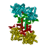
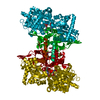
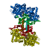
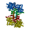
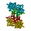
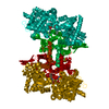
 Components Components | GLYCOGEN PHOSPHORYLASE, MUSCLE FORM | ||||||
 Keywords Keywords | TRANSFERASE / TYPE 2 DIABETES / INHIBITOR / STRUCTURE-BASED DRUG DESIGN | ||||||
| Function / homology |  Function and homology information Function and homology informationglycogen phosphorylase / glycogen phosphorylase activity / glycogen catabolic process / skeletal muscle myofibril / pyridoxal phosphate binding / nucleotide binding Similarity search - Function | ||||||
| Biological species |  | ||||||
| Method |  X-RAY DIFFRACTION / SYNCHROTRON / MOLECULAR REPLACEMENT / Resolution: 1.95 Å X-RAY DIFFRACTION / SYNCHROTRON / MOLECULAR REPLACEMENT / Resolution: 1.95 Å | ||||||
 Authors Authors | Alexacou, K.M. / Papakonstantinou, M. / Leonidas, D.D. / Zographos, S.E. / Chrysina, E.D. | ||||||
 Citation Citation | Journal: Bioorg.Med.Chem. / Year: 2014 Title: Glucopyranosylidene-Spiro-Iminothiazolidinone, a New Bicyclic Ring System: Synthesis, Derivatization, and Evaluation for Inhibition of Glycogen Phosphorylase by Enzyme Kinetic and Crystallographic Methods. Authors: Czifrak, K. / Deak, S. / Pahi, A. / Kover, K.E. / Docsa, T. / Gergely, P. / Alexacou, K.M. / Papakonstantinou, M. / Leonidas, D.D. / Zographos, S.E. / Chrysina, E.D. / Somsak, L. #1: Journal: Protein Sci. / Year: 1998Title: The Structure of a Glycogen Phosphorylase Glucopyranose Spirohydantoin Complex at 1.8 A Resolution and 100 K: The Role of the Water Structure and its Contribution to Binding. Authors: Gregoriou, M. / Noble, M.E. / Watson, K.A. / Garman, E.F. / Krulle, T.M. / De La Fuente, C. / Fleet, G.W. / Oikonomakos, N.G. / Johnson, L.N. | ||||||
| History |
|
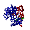
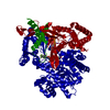
- Structure visualization
| Structure viewer | Molecule: MolmilJmol/JSmol |
|---|
- Downloads & links
-Download
| PDBx/mmCIF format | 4ctm.cif.gz | 180.6 KB | Display | PDBx/mmCIF format |
|---|---|---|---|---|
| PDB format | pdb4ctm.ent.gz | 141.2 KB | Display | PDB format |
| PDBx/mmJSON format | 4ctm.json.gz | Tree view | PDBx/mmJSON format | |
| Others |  Other downloads Other downloads |
-Validation report
| Arichive directory | https://data.pdbj.org/pub/pdb/validation_reports/ct/4ctmftp://data.pdbj.org/pub/pdb/validation_reports/ct/4ctm | HTTPS FTP |
|---|
-Related structure data
| Related structure data |  4ctnC  4ctoC  2pydS C: citing same article ( S: Starting model for refinement |
|---|---|
| Similar structure data |


-Links
 PDBj
PDBj
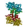
- Assembly
| Deposited unit | 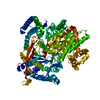
| ||||||||
|---|---|---|---|---|---|---|---|---|---|
| 1 | 
| ||||||||
| Unit cell |
|
-Components
| #1: Protein | Mass: 97422.398 Da / Num. of mol.: 1 / Source method: isolated from a natural source / Source: (natural) |
|---|---|
| #2: Chemical | ChemComp-MIF / (  Mass: 264.256 Da / Num. of mol.: 1 / Source method: obtained synthetically / Formula: C8H12N2O6S Mass: 264.256 Da / Num. of mol.: 1 / Source method: obtained synthetically / Formula: C8H12N2O6S |
| #3: Chemical | ChemComp-PLP /   Mass: 247.142 Da / Num. of mol.: 1 / Source method: obtained synthetically / Formula: C8H10NO6P Mass: 247.142 Da / Num. of mol.: 1 / Source method: obtained synthetically / Formula: C8H10NO6P |
| #4: Chemical | ChemComp-PO4 /   Mass: 94.971 Da / Num. of mol.: 1 / Source method: obtained synthetically / Formula: PO4 Mass: 94.971 Da / Num. of mol.: 1 / Source method: obtained synthetically / Formula: PO4 |
| #5: Water | ChemComp-HOH /  Mass: 18.015 Da / Num. of mol.: 274 / Source method: isolated from a natural source / Formula: H2O Mass: 18.015 Da / Num. of mol.: 274 / Source method: isolated from a natural source / Formula: H2O |
-Experimental details
-Experiment
| Experiment | Method: X-RAY DIFFRACTION / Number of used crystals: 1 |
|---|
- Sample preparation
| Crystal | Density Matthews: 2.46 Å3/Da / Density % sol: 49.95 % / Description: NONE |
|---|---|
| Crystal grow | pH: 6.8 Details: 25MG/ML ENZYME, 1MM SPERMINE, 10MM BES, 3MM DTT, 0.1MM EDTA, 0.02% SODIUM AZIDE, PH 6.7 |
-Data collection
| Diffraction | Mean temperature: 298 K |
|---|---|
| Diffraction source | Source: SYNCHROTRON / Site: SRS  / Beamline: PX10.1 / Wavelength: 0.97976 / Beamline: PX10.1 / Wavelength: 0.97976 |
| Detector | Type: MARRESEARCH / Detector: CCD / Date: Nov 16, 2007 / Details: MIRRORS |
| Radiation | Monochromator: GRAPHITE / Protocol: SINGLE WAVELENGTH / Monochromatic (M) / Laue (L): M / Scattering type: x-ray |
| Radiation wavelength | Wavelength: 0.97976 Å / Relative weight: 1 |
| Reflection | Resolution: 1.95→35.7 Å / Num. obs: 69137 / % possible obs: 98 % / Observed criterion σ(I): 0 / Redundancy: 5.3 % / Biso Wilson estimate: 25.4 Å2 / Rmerge(I) obs: 0.06 / Net I/σ(I): 20.4 |
| Reflection shell | Resolution: 1.95→2.06 Å / Redundancy: 5.2 % / Rmerge(I) obs: 0.38 / Mean I/σ(I) obs: 5.1 / % possible all: 96.8 |
- Processing
| Software |
| ||||||||||||||||||||||||||||||||||||||||||||||||||||||||||||||||||||||||||||||||||||||||||||||||||||||||||||||||||||||||||||||||||||||||||||||||||||||||||||||||||||||||||||||||||||||
|---|---|---|---|---|---|---|---|---|---|---|---|---|---|---|---|---|---|---|---|---|---|---|---|---|---|---|---|---|---|---|---|---|---|---|---|---|---|---|---|---|---|---|---|---|---|---|---|---|---|---|---|---|---|---|---|---|---|---|---|---|---|---|---|---|---|---|---|---|---|---|---|---|---|---|---|---|---|---|---|---|---|---|---|---|---|---|---|---|---|---|---|---|---|---|---|---|---|---|---|---|---|---|---|---|---|---|---|---|---|---|---|---|---|---|---|---|---|---|---|---|---|---|---|---|---|---|---|---|---|---|---|---|---|---|---|---|---|---|---|---|---|---|---|---|---|---|---|---|---|---|---|---|---|---|---|---|---|---|---|---|---|---|---|---|---|---|---|---|---|---|---|---|---|---|---|---|---|---|---|---|---|---|---|
| Refinement | Method to determine structure: MOLECULAR REPLACEMENT Starting model: PDB ENTRY 2PYD Resolution: 1.95→35.71 Å / Cor.coef. Fo:Fc: 0.967 / Cor.coef. Fo:Fc free: 0.958 / SU B: 2.873 / SU ML: 0.083 / Cross valid method: THROUGHOUT / ESU R: 0.147 / ESU R Free: 0.127 / Stereochemistry target values: MAXIMUM LIKELIHOOD / Details: HYDROGENS HAVE BEEN ADDED IN THE RIDING POSITIONS.
| ||||||||||||||||||||||||||||||||||||||||||||||||||||||||||||||||||||||||||||||||||||||||||||||||||||||||||||||||||||||||||||||||||||||||||||||||||||||||||||||||||||||||||||||||||||||
| Solvent computation | Ion probe radii: 0.8 Å / Shrinkage radii: 0.8 Å / VDW probe radii: 1.2 Å / Solvent model: MASK | ||||||||||||||||||||||||||||||||||||||||||||||||||||||||||||||||||||||||||||||||||||||||||||||||||||||||||||||||||||||||||||||||||||||||||||||||||||||||||||||||||||||||||||||||||||||
| Displacement parameters | Biso mean: 31.349 Å2 | ||||||||||||||||||||||||||||||||||||||||||||||||||||||||||||||||||||||||||||||||||||||||||||||||||||||||||||||||||||||||||||||||||||||||||||||||||||||||||||||||||||||||||||||||||||||
| Refinement step | Cycle: LAST / Resolution: 1.95→35.71 Å
| ||||||||||||||||||||||||||||||||||||||||||||||||||||||||||||||||||||||||||||||||||||||||||||||||||||||||||||||||||||||||||||||||||||||||||||||||||||||||||||||||||||||||||||||||||||||
| Refine LS restraints |
|