 Movie
Movie Controller

[English] 日本語
 Yorodumi
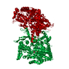
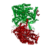
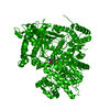
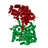
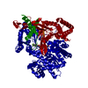
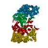
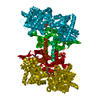
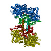
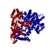
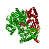
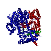
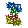
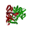
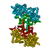
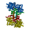
- PDB-2gpb: COMPARISON OF THE BINDING OF GLUCOSE AND GLUCOSE-1-PHOSPHATE DERI... -
+ Open data

- Basic information
| Entry | Database: PDB / ID: 2gpb | ||||||
|---|---|---|---|---|---|---|---|
| Title | COMPARISON OF THE BINDING OF GLUCOSE AND GLUCOSE-1-PHOSPHATE DERIVATIVES TO T-STATE GLYCOGEN PHOSPHORYLASE B | ||||||
 Components Components | GLYCOGEN PHOSPHORYLASE B | ||||||
 Keywords Keywords | GLYCOGEN PHOSPHORYLASE | ||||||
| Function / homology |  Function and homology information Function and homology informationglycogen phosphorylase / glycogen phosphorylase activity / glycogen catabolic process / skeletal muscle myofibril / pyridoxal phosphate binding / nucleotide binding Similarity search - Function | ||||||
| Biological species |  | ||||||
| Method |  X-RAY DIFFRACTION / Resolution: 2.3 Å X-RAY DIFFRACTION / Resolution: 2.3 Å | ||||||
 Authors Authors | Martin, J.L. / Johnson, L.N. | ||||||
 Citation Citation | Journal: Biochemistry / Year: 1990 Title: Comparison of the binding of glucose and glucose 1-phosphate derivatives to T-state glycogen phosphorylase b. Authors: Martin, J.L. / Johnson, L.N. / Withers, S.G. #1: Journal: Glycogen Phosphorylase B: Description of the Protein StructureTitle: Glycogen Phosphorylase B: Description of the Protein Structure 1 1991 Authors: Acharya, K.R. / Stuart, D.I. / Varvill, K.M. / Johnson, L.N. #2: Journal: J.Mol.Biol. / Year: 1991Title: Structural Mechanism for Glycogen Phosphorylase Control by Phosphorylation and AMP Authors: Barford, D. / Hu, S.-H. / Johnson, L.N. #3: Journal: J.Mol.Biol. / Year: 1990Title: Refined Crystal Structure of the Phosphorylase-Heptulose 2-Phosphate-Oligosaccharide-AMP Complex Authors: Johnson, L.N. / Acharya, K.R. / Jordan, M.D. / Mclaughlin, P.J. #4: Journal: Nature / Year: 1989Title: The Allosteric Transition of Glycogen Phosphorylase Authors: Barford, D. / Johnson, L.N. #5: Journal: Nature / Year: 1988Title: Structural Changes in Glycogen Phosphorylase Induced by Phosphorylation Authors: Sprang, S.R. / Acharya, K.R. / Goldsmith, E.J. / Stuart, D.I. / Varvill, K. / Fletterick, R.J. / Madsen, N.B. / Johnson, L.N. | ||||||
| History |
|
- Structure visualization
| Structure viewer | Molecule: MolmilJmol/JSmol |
|---|
- Downloads & links
-Download
| PDBx/mmCIF format | 2gpb.cif.gz | 195.4 KB | Display | PDBx/mmCIF format |
|---|---|---|---|---|
| PDB format | pdb2gpb.ent.gz | 152.5 KB | Display | PDB format |
| PDBx/mmJSON format | 2gpb.json.gz | Tree view | PDBx/mmJSON format | |
| Others |  Other downloads Other downloads |
-Validation report
| Arichive directory | https://data.pdbj.org/pub/pdb/validation_reports/gp/2gpbftp://data.pdbj.org/pub/pdb/validation_reports/gp/2gpb | HTTPS FTP |
|---|
-Related structure data


-Links
 PDBj
PDBj


- Assembly
| Deposited unit | 
| ||||||||
|---|---|---|---|---|---|---|---|---|---|
| 1 | 
| ||||||||
| Unit cell |
| ||||||||
| Atom site foot note | 1: RESIDUE 380 IS LEU IN THE SEQUENCE (K.NAKANO,P.K.HWANG, R.J.FLETTERICK, FEBS LETT., V. 204, P. 283, 1986) BUT IT HAS BEEN PRESENTED AS ILE IN THIS ENTRY. THIS ASSIGNMENT WAS MADE IN THE ORIGINAL ...1: RESIDUE 380 IS LEU IN THE SEQUENCE (K.NAKANO,P.K.HWANG, R.J.FLETTERICK, FEBS LETT., V. 204, P. 283, 1986) BUT IT HAS BEEN PRESENTED AS ILE IN THIS ENTRY. THIS ASSIGNMENT WAS MADE IN THE ORIGINAL STRUCTURE DETERMINATION AT 1.9 ANGSTROMS (PRESENTED IN PROTEIN DATA BANK ENTRY 1GPB) AND CARRIED THROUGH TO THE OTHER ENTRIES. ILE IS MORE CONSISTENT WITH THE ELECTRON DENSITY. HOWEVER, THE RESOLUTION AT 1.9 ANGSTROMS DOES NOT ALLOW A DEFINITIVE ASSIGNMENT. |
-Components
| #1: Protein | Mass: 97291.203 Da / Num. of mol.: 1 Source method: isolated from a genetically manipulated source Source: (gene. exp.) |
|---|---|
| #2: Sugar | ChemComp-GLC /   Type: D-saccharide, alpha linking / Mass: 180.156 Da / Num. of mol.: 1 Type: D-saccharide, alpha linking / Mass: 180.156 Da / Num. of mol.: 1Source method: isolated from a genetically manipulated source Formula: C6H12O6 |
| #3: Chemical | ChemComp-PLP /   Mass: 247.142 Da / Num. of mol.: 1 / Source method: obtained synthetically / Formula: C8H10NO6P Mass: 247.142 Da / Num. of mol.: 1 / Source method: obtained synthetically / Formula: C8H10NO6P |
| #4: Water | ChemComp-HOH /  Mass: 18.015 Da / Num. of mol.: 575 / Source method: isolated from a natural source / Formula: H2O Mass: 18.015 Da / Num. of mol.: 575 / Source method: isolated from a natural source / Formula: H2O |
| Has protein modification | N |
| Sequence details | RESIDUE 380 IS LEU IN THE SEQUENCE (K.NAKANO,P.K.HWANG, R.J.FLETTERICK, FEBS LETT., V. 204, P. 283, ...RESIDUE 380 IS LEU IN THE SEQUENCE (K.NAKANO,P.K.HWANG, R.J.FLETTERICK |
-Experimental details
-Experiment
| Experiment | Method: X-RAY DIFFRACTION |
|---|
- Sample preparation
| Crystal | Density Matthews: 2.47 Å3/Da / Density % sol: 50.13 % | |||||||||||||||||||||||||||||||||||
|---|---|---|---|---|---|---|---|---|---|---|---|---|---|---|---|---|---|---|---|---|---|---|---|---|---|---|---|---|---|---|---|---|---|---|---|---|
| Crystal grow | *PLUS Temperature: unknown K / pH: 6.7 / Method: unknown | |||||||||||||||||||||||||||||||||||
| Components of the solutions | *PLUS
|
-Data collection
| Reflection | *PLUS Highest resolution: 2.3 Å / Num. obs: 34771 / % possible obs: 82 % / Num. measured all: 85168 / Rmerge(I) obs: 0.069 |
|---|
- Processing
| Software | Name: X-PLOR / Classification: refinement | ||||||||||||||||||||||||||||||||||||||||||||||||||||||||||||
|---|---|---|---|---|---|---|---|---|---|---|---|---|---|---|---|---|---|---|---|---|---|---|---|---|---|---|---|---|---|---|---|---|---|---|---|---|---|---|---|---|---|---|---|---|---|---|---|---|---|---|---|---|---|---|---|---|---|---|---|---|---|
| Refinement | Resolution: 2.3→8 Å /
| ||||||||||||||||||||||||||||||||||||||||||||||||||||||||||||
| Refinement step | Cycle: LAST / Resolution: 2.3→8 Å
| ||||||||||||||||||||||||||||||||||||||||||||||||||||||||||||
| Refine LS restraints |
| ||||||||||||||||||||||||||||||||||||||||||||||||||||||||||||
| Software | *PLUS Name: X-PLOR / Classification: refinement | ||||||||||||||||||||||||||||||||||||||||||||||||||||||||||||
| Refinement | *PLUS Rfactor Rwork: 0.181 | ||||||||||||||||||||||||||||||||||||||||||||||||||||||||||||
| Solvent computation | *PLUS | ||||||||||||||||||||||||||||||||||||||||||||||||||||||||||||
| Displacement parameters | *PLUS | ||||||||||||||||||||||||||||||||||||||||||||||||||||||||||||
| Refine LS restraints | *PLUS Type: x_angle_d / Dev ideal: 3.4 |
