[English] 日本語
Yorodumi
- PDB-6f3l: The crystal structure of Glycogen Phosphorylase in complex with 10b -

+
Open data


ID or keywords:

Loading...

-
Basic information

Entry
Database: PDB / ID: 6f3l
TitleThe crystal structure of Glycogen Phosphorylase in complex with 10b
ComponentsGlycogen phosphorylase, muscle form
KeywordsTRANSFERASE
Function / homology
Function and homology information


glycogen phosphorylase / glycogen phosphorylase activity / linear malto-oligosaccharide phosphorylase activity / SHG alpha-glucan phosphorylase activity / glycogen catabolic process / skeletal muscle myofibril / pyridoxal phosphate binding / nucleotide binding
Similarity search - Function
Glycosyl transferase, family 35 / Glycogen/starch/alpha-glucan phosphorylase / Phosphorylase pyridoxal-phosphate attachment site / Carbohydrate phosphorylase / Phosphorylase pyridoxal-phosphate attachment site. / Glycogen Phosphorylase B; / Rossmann fold / 3-Layer(aba) Sandwich / Alpha Beta
Similarity search - Domain/homology
Chem-CJW / PYRIDOXAL-5'-PHOSPHATE / Glycogen phosphorylase, muscle form
Similarity search - Component
Biological speciesOryctolagus cuniculus (rabbit)
MethodX-RAY DIFFRACTION / SYNCHROTRON / Resolution: 1.9 Å
AuthorsKyriakis, E. / Barkas, T.A. / Stravodimos, G.A. / Skamnaki, V.T. / Leonidas, D.D.
CitationJournal: Eur J Med Chem / Year: 2018
Title: A multidisciplinary study of 3-( beta-d-glucopyranosyl)-5-substituted-1,2,4-triazole derivatives as glycogen phosphorylase inhibitors: Computation, synthesis, crystallography and kinetics ...Title: A multidisciplinary study of 3-( beta-d-glucopyranosyl)-5-substituted-1,2,4-triazole derivatives as glycogen phosphorylase inhibitors: Computation, synthesis, crystallography and kinetics reveal new potent inhibitors.
Authors: Kun, S. / Begum, J. / Kyriakis, E. / Stamati, E.C.V. / Barkas, T.A. / Szennyes, E. / Bokor, E. / Szabo, K.E. / Stravodimos, G.A. / Sipos, A. / Docsa, T. / Gergely, P. / Moffatt, C. / ...Authors: Kun, S. / Begum, J. / Kyriakis, E. / Stamati, E.C.V. / Barkas, T.A. / Szennyes, E. / Bokor, E. / Szabo, K.E. / Stravodimos, G.A. / Sipos, A. / Docsa, T. / Gergely, P. / Moffatt, C. / Patraskaki, M.S. / Kokolaki, M.C. / Gkerdi, A. / Skamnaki, V.T. / Leonidas, D.D. / Somsak, L. / Hayes, J.M.
History
DepositionNov 28, 2017Deposition site: PDBE / Processing site: PDBE
Revision 1.0Feb 28, 2018Provider: repository / Type: Initial release

-
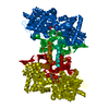
Structure visualization

Structure viewerMolecule:
MolmilJmol/JSmol

Downloads & links

-
Assembly

Deposited unit
A: Glycogen phosphorylase, muscle form
hetero molecules


Theoretical massNumber of molelcules
Total (without water)98,0713
Polymers97,4221
Non-polymers6492
Water4,504250
1
A: Glycogen phosphorylase, muscle form
hetero molecules

A: Glycogen phosphorylase, muscle form
hetero molecules


Theoretical massNumber of molelcules
Total (without water)196,1426
Polymers194,8452
Non-polymers1,2974
Water362
TypeNameSymmetry operationNumber
identity operation1_555x,y,z1
crystal symmetry operation7_556y,x,-z+11
Buried area5770 Å2
ΔGint-22 kcal/mol
Surface area57270 Å2
MethodPISA
Unit cell
Length a, b, c (Å)128.646, 128.646, 116.276
Angle α, β, γ (deg.)90.00, 90.00, 90.00
Int Tables number96
Space group name H-MP43212

-
Components

#1: Protein Glycogen phosphorylase, muscle form / / Myophosphorylase


Mass: 97422.398 Da / Num. of mol.: 1 / Source method: isolated from a natural source / Source: (natural) Oryctolagus cuniculus (rabbit) / Tissue: muscleSkeletal muscle / References: UniProt: P00489, glycogen phosphorylase
#2: Chemical ChemComp-PLP / PYRIDOXAL-5'-PHOSPHATE / VITAMIN B6 Phosphate / Pyridoxal phosphate


Mass: 247.142 Da / Num. of mol.: 1 / Source method: obtained synthetically / Formula: C8H10NO6P
#3: Chemical ChemComp-CJW / 6-[5-[(2~{S},3~{R},4~{R},5~{S},6~{R})-6-(hydroxymethyl)-3,4,5-tris(oxidanyl)oxan-2-yl]-1~{H}-1,2,4-triazol-3-yl]naphthalene-2-carboxylic acid


Mass: 401.370 Da / Num. of mol.: 1 / Source method: obtained synthetically / Formula: C19H19N3O7
#4: Water ChemComp-HOH / water / Water


Mass: 18.015 Da / Num. of mol.: 250 / Source method: isolated from a natural source / Formula: H2O

-
Experimental details

-
Experiment

ExperimentMethod: X-RAY DIFFRACTION / Number of used crystals: 1

-
Sample preparation

CrystalDensity Matthews: 2.47 Å3/Da / Density % sol: 50.18 %
Crystal growTemperature: 289 K / Method: small tubes / pH: 6.7 / Details: 10 mM BES buffer

-
Data collection

DiffractionMean temperature: 293 K
Diffraction sourceSource: SYNCHROTRON / Site: Diamond / Beamline: I04 / Wavelength: 0.9795 Å
DetectorType: DECTRIS PILATUS 6M-F / Detector: PIXEL / Date: Oct 13, 2017
RadiationProtocol: SINGLE WAVELENGTH / Monochromatic (M) / Laue (L): M / Scattering type: x-ray
Radiation wavelengthWavelength: 0.9795 Å / Relative weight: 1
ReflectionResolution: 1.9→129 Å / Num. obs: 76655 / % possible obs: 99.5 % / Redundancy: 14.9 % / Rmerge(I) obs: 0.163 / Net I/σ(I): 10.8
Reflection shellResolution: 1.9→1.94 Å / Redundancy: 15.3 % / Mean I/σ(I) obs: 3.4 / % possible all: 98.9

-
Processing

Software
NameVersionClassification
REFMAC5.8.0158refinement
MOSFLMdata reduction
Aimlessdata scaling
RefinementResolution: 1.9→129 Å / Cor.coef. Fo:Fc: 0.978 / Cor.coef. Fo:Fc free: 0.964 / SU B: 4.692 / SU ML: 0.067 / Cross valid method: THROUGHOUT / ESU R: 0.104 / ESU R Free: 0.096 / Details: HYDROGENS HAVE BEEN ADDED IN THE RIDING POSITIONS
RfactorNum. reflection% reflectionSelection details
Rfree0.16146 3768 4.9 %RANDOM
Rwork0.13709 ---
obs0.13829 72835 99.36 %-
Solvent computationIon probe radii: 0.7 Å / Shrinkage radii: 0.7 Å / VDW probe radii: 1 Å
Displacement parametersBiso mean: 41.04 Å2
Baniso -1Baniso -2Baniso -3
1-0.97 Å2-0 Å2-0 Å2
2--0.97 Å2-0 Å2
3----1.93 Å2
Refinement stepCycle: 1 / Resolution: 1.9→129 Å
ProteinNucleic acidLigandSolventTotal
Num. atoms6589 0 44 250 6883
Refine LS restraints
Refine-IDTypeDev idealDev ideal targetNumber
X-RAY DIFFRACTIONr_bond_refined_d0.010.0196825
X-RAY DIFFRACTIONr_bond_other_d0.0010.026320
X-RAY DIFFRACTIONr_angle_refined_deg1.3111.9539254
X-RAY DIFFRACTIONr_angle_other_deg0.9462.99714612
X-RAY DIFFRACTIONr_dihedral_angle_1_deg5.785821
X-RAY DIFFRACTIONr_dihedral_angle_2_deg34.14423.561351
X-RAY DIFFRACTIONr_dihedral_angle_3_deg12.787151183
X-RAY DIFFRACTIONr_dihedral_angle_4_deg17.3111560
X-RAY DIFFRACTIONr_chiral_restr0.0820.21000
X-RAY DIFFRACTIONr_gen_planes_refined0.0060.0217604
X-RAY DIFFRACTIONr_gen_planes_other0.0010.021471
X-RAY DIFFRACTIONr_nbd_refined
X-RAY DIFFRACTIONr_nbd_other
X-RAY DIFFRACTIONr_nbtor_refined
X-RAY DIFFRACTIONr_nbtor_other
X-RAY DIFFRACTIONr_xyhbond_nbd_refined
X-RAY DIFFRACTIONr_xyhbond_nbd_other
X-RAY DIFFRACTIONr_metal_ion_refined
X-RAY DIFFRACTIONr_metal_ion_other
X-RAY DIFFRACTIONr_symmetry_vdw_refined
X-RAY DIFFRACTIONr_symmetry_vdw_other
X-RAY DIFFRACTIONr_symmetry_hbond_refined
X-RAY DIFFRACTIONr_symmetry_hbond_other
X-RAY DIFFRACTIONr_symmetry_metal_ion_refined
X-RAY DIFFRACTIONr_symmetry_metal_ion_other
X-RAY DIFFRACTIONr_mcbond_it2.0962.5343251
X-RAY DIFFRACTIONr_mcbond_other2.0962.5333250
X-RAY DIFFRACTIONr_mcangle_it2.9793.7834062
X-RAY DIFFRACTIONr_mcangle_other2.9793.7844063
X-RAY DIFFRACTIONr_scbond_it3.4393.083574
X-RAY DIFFRACTIONr_scbond_other3.4393.083573
X-RAY DIFFRACTIONr_scangle_it
X-RAY DIFFRACTIONr_scangle_other5.4244.435186
X-RAY DIFFRACTIONr_long_range_B_refined6.96329.7967518
X-RAY DIFFRACTIONr_long_range_B_other6.96229.7957519
X-RAY DIFFRACTIONr_rigid_bond_restr
X-RAY DIFFRACTIONr_sphericity_free
X-RAY DIFFRACTIONr_sphericity_bonded
LS refinement shellResolution: 1.9→1.949 Å / Total num. of bins used: 20
RfactorNum. reflection% reflection
Rfree0.219 272 -
Rwork0.181 5298 -
obs--98.88 %
Refinement TLS params.Method: refined / Origin x: 28.276 Å / Origin y: 21.363 Å / Origin z: 31.601 Å
111213212223313233
T0.0377 Å2-0.05 Å20.0051 Å2-0.0859 Å2-0.033 Å2--0.0413 Å2
L0.6088 °20.0678 °2-0.0311 °2-0.4987 °2-0.1376 °2--0.922 °2
S-0.0427 Å °0.0483 Å °0.0569 Å °-0.0222 Å °0.0075 Å °0.0161 Å °0.0788 Å °-0.1068 Å °0.0352 Å °

+
About Yorodumi

-
News

-
Feb 9, 2022. New format data for meta-information of EMDB entries

New format data for meta-information of EMDB entries

  • Version 3 of the EMDB header file is now the official format.
  • The previous official version 1.9 will be removed from the archive.

Related info.:EMDB header

External links:wwPDB to switch to version 3 of the EMDB data model

-
Aug 12, 2020. Covid-19 info

Covid-19 info

URL: https://pdbj.org/emnavi/covid19.php

New page: Covid-19 featured information page in EM Navigator.

Related info.:Covid-19 info / Mar 5, 2020. Novel coronavirus structure data

+
Mar 5, 2020. Novel coronavirus structure data

Novel coronavirus structure data

Related info.:Yorodumi Speices / Aug 12, 2020. Covid-19 info

External links:COVID-19 featured content - PDBj / Molecule of the Month (242):Coronavirus Proteases

+
Jan 31, 2019. EMDB accession codes are about to change! (news from PDBe EMDB page)

EMDB accession codes are about to change! (news from PDBe EMDB page)

  • The allocation of 4 digits for EMDB accession codes will soon come to an end. Whilst these codes will remain in use, new EMDB accession codes will include an additional digit and will expand incrementally as the available range of codes is exhausted. The current 4-digit format prefixed with “EMD-” (i.e. EMD-XXXX) will advance to a 5-digit format (i.e. EMD-XXXXX), and so on. It is currently estimated that the 4-digit codes will be depleted around Spring 2019, at which point the 5-digit format will come into force.
  • The EM Navigator/Yorodumi systems omit the EMD- prefix.

Related info.:Q: What is EMD? / ID/Accession-code notation in Yorodumi/EM Navigator

External links:EMDB Accession Codes are Changing Soon! / Contact to PDBj

+
Jul 12, 2017. Major update of PDB

Major update of PDB

  • wwPDB released updated PDB data conforming to the new PDBx/mmCIF dictionary.
  • This is a major update changing the version number from 4 to 5, and with Remediation, in which all the entries are updated.
  • In this update, many items about electron microscopy experimental information are reorganized (e.g. em_software).
  • Now, EM Navigator and Yorodumi are based on the updated data.

External links:wwPDB Remediation / Enriched Model Files Conforming to OneDep Data Standards Now Available in the PDB FTP Archive

-
Yorodumi

Thousand views of thousand structures

  • Yorodumi is a browser for structure data from EMDB, PDB, SASBDB, etc.
  • This page is also the successor to EM Navigator detail page, and also detail information page/front-end page for Omokage search.
  • The word "yorodu" (or yorozu) is an old Japanese word meaning "ten thousand". "mi" (miru) is to see.

Related info.:EMDB / PDB / SASBDB / Comparison of 3 databanks / Yorodumi Search / Aug 31, 2016. New EM Navigator & Yorodumi / Yorodumi Papers / Jmol/JSmol / Function and homology information / Changes in new EM Navigator and Yorodumi

Read more