- PDB-6l8o: Crystal structure of the K. lactis Rad5 (Hg-derivative) -
+
Open data
ID or keywords:
Loading...
-
Basic information
Entry
Database: PDB / ID: 6l8o
Title
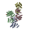
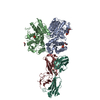
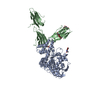
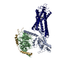
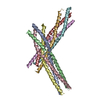
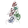
Crystal structure of the K. lactis Rad5 (Hg-derivative)
Components
DNA repair protein RAD5
Keywords
DNA BINDING PROTEIN / DNA damage tolerance / Helicase / Snf2 family
Function / homology
Function and homology information
ATP-dependent activity, acting on DNA / helicase activity / RING-type E3 ubiquitin transferase / Hydrolases; Acting on acid anhydrides; Acting on acid anhydrides to facilitate cellular and subcellular movement / ubiquitin protein ligase activity / single-stranded DNA binding / double-stranded DNA binding / DNA repair / ATP hydrolysis activity / zinc ion binding ...ATP-dependent activity, acting on DNA / helicase activity / RING-type E3 ubiquitin transferase / Hydrolases; Acting on acid anhydrides; Acting on acid anhydrides to facilitate cellular and subcellular movement / ubiquitin protein ligase activity / single-stranded DNA binding / double-stranded DNA binding / DNA repair / ATP hydrolysis activity / zinc ion binding / ATP binding / nucleus / cytoplasm Similarity search - Function
In the structure databanks used in Yorodumi, some data are registered as the other names, "COVID-19 virus" and "2019-nCoV". Here are the details of the virus and the list of structure data.
Jan 31, 2019. EMDB accession codes are about to change! (news from PDBe EMDB page)
EMDB accession codes are about to change! (news from PDBe EMDB page)
The allocation of 4 digits for EMDB accession codes will soon come to an end. Whilst these codes will remain in use, new EMDB accession codes will include an additional digit and will expand incrementally as the available range of codes is exhausted. The current 4-digit format prefixed with “EMD-” (i.e. EMD-XXXX) will advance to a 5-digit format (i.e. EMD-XXXXX), and so on. It is currently estimated that the 4-digit codes will be depleted around Spring 2019, at which point the 5-digit format will come into force.
The EM Navigator/Yorodumi systems omit the EMD- prefix.
Related info.:Q: What is EMD? / ID/Accession-code notation in Yorodumi/EM Navigator
Yorodumi is a browser for structure data from EMDB, PDB, SASBDB, etc.
This page is also the successor to EM Navigator detail page, and also detail information page/front-end page for Omokage search.
The word "yorodu" (or yorozu) is an old Japanese word meaning "ten thousand". "mi" (miru) is to see.
Related info.:EMDB / PDB / SASBDB / Comparison of 3 databanks / Yorodumi Search / Aug 31, 2016. New EM Navigator & Yorodumi / Yorodumi Papers / Jmol/JSmol / Function and homology information / Changes in new EM Navigator and Yorodumi
 Movie
Movie Controller


 Open data
 Basic information
 Components
 Keywords
 Function and homology information
 Kluyveromyces lactis NRRL Y-1140 (yeast)
 X-RAY DIFFRACTION /
  Authors
 China, 1items
  Citation
 Structure visualization
 Downloads & links
 Other downloads
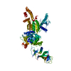


 PDBj
PDBj


 Assembly
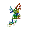


 Mass: 200.590 Da / Num. of mol.: 3 / Source method: obtained synthetically / Formula: Hg
 Mass: 122.143 Da / Num. of mol.: 1 / Source method: obtained synthetically / Formula: C4H12NO3 / Comment: pH buffer*YM
 Sample preparation
 Processing