National Institutes of Health/National Institute Of Allergy and Infectious Diseases (NIH/NIAID)
AI039657
United States
National Institutes of Health/National Institute Of Allergy and Infectious Diseases (NIH/NIAID)
AI118932
United States
National Institutes of Health/National Cancer Institute (NIH/NCI)
CA116087
United States
National Institutes of Health/Office of the Director
S10OD030275
United States
National Institutes of Health/National Institute of General Medical Sciences (NIH/NIGMS)
T32GM08320
United States
National Institutes of Health/National Institute of General Medical Sciences (NIH/NIGMS)
T32GM007315
United States
National Institutes of Health/National Institute of General Medical Sciences (NIH/NIGMS)
F31GM139291
United States
American Heart Association
905705
United States
Department of Veterans Affairs (VA, United States)
5I01BX004447
United States
Citation
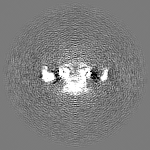
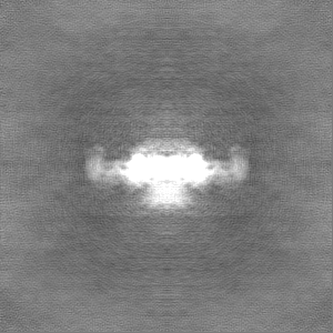
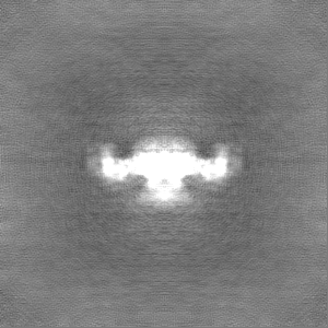
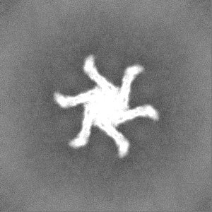
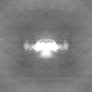
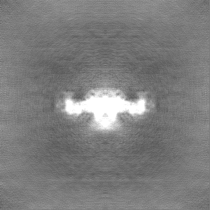
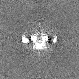
Journal: J Mol Biol / Year: 2024 Title: Structural Analysis of Membrane-associated Forms of Helicobacter pylori VacA Toxin. Authors: Sarah M Connolly / Amanda L Erwin / Megan Sabb / Jessica L Hanks / Louise Chang / Rachel M Torrez / Georgia C Caso / Anne M Campbell / Shyamal Mosalaganti / Timothy L Cover / Melanie D Ohi / Abstract: Helicobacter pylori colonizes the stomach in about half of the human population, leading to an increased risk of peptic ulcer disease and gastric cancer. H. pylori secretes an 88 kDa VacA toxin that ...Helicobacter pylori colonizes the stomach in about half of the human population, leading to an increased risk of peptic ulcer disease and gastric cancer. H. pylori secretes an 88 kDa VacA toxin that contributes to pathogenesis. VacA assembles into oligomeric complexes in solution and forms anion-selective channels in cell membranes. Cryo-electron microscopy (cryo-EM) analyses of VacA oligomers in solution provided insights into VacA oligomerization but failed to reveal the structure of the hydrophobic N-terminal region predicted to be a pore-forming domain. In this study, we incubated VacA with liposomes and used single particle cryo-EM to analyze detergent-extracted VacA oligomers. A 3D structure of detergent-solubilized VacA hexamers revealed the presence of six α-helices extending from the center of the oligomers, a feature not observed in previous studies of water-soluble VacA oligomers. Cryo-electron tomography analysis and 2D averages of VacA associated with liposomes confirmed that central regions of the membrane-associated VacA oligomers can insert into the lipid bilayer. However, insertion is heterogenous, with some membrane-associated oligomers appearing only partially inserted and others sitting on top of the bilayer. These studies indicate that VacA undergoes a conformational change when contacting the membrane and reveal an α-helical region positioned to extend into the membrane. Although the reported VacA 3D structure does not represent a selective anion channel, our combined single particle 3D analysis, cryo-electron tomography, and modeling allow us to propose a model for the structural organization of the VacA N-terminus in the context of a hexamer as it inserts into the membrane.
In the structure databanks used in Yorodumi, some data are registered as the other names, "COVID-19 virus" and "2019-nCoV". Here are the details of the virus and the list of structure data.
Jan 31, 2019. EMDB accession codes are about to change! (news from PDBe EMDB page)
EMDB accession codes are about to change! (news from PDBe EMDB page)
The allocation of 4 digits for EMDB accession codes will soon come to an end. Whilst these codes will remain in use, new EMDB accession codes will include an additional digit and will expand incrementally as the available range of codes is exhausted. The current 4-digit format prefixed with “EMD-” (i.e. EMD-XXXX) will advance to a 5-digit format (i.e. EMD-XXXXX), and so on. It is currently estimated that the 4-digit codes will be depleted around Spring 2019, at which point the 5-digit format will come into force.
The EM Navigator/Yorodumi systems omit the EMD- prefix.
Related info.:Q: What is EMD? / ID/Accession-code notation in Yorodumi/EM Navigator
Yorodumi is a browser for structure data from EMDB, PDB, SASBDB, etc.
This page is also the successor to EM Navigator detail page, and also detail information page/front-end page for Omokage search.
The word "yorodu" (or yorozu) is an old Japanese word meaning "ten thousand". "mi" (miru) is to see.
Related info.:EMDB / PDB / SASBDB / Comparison of 3 databanks / Yorodumi Search / Aug 31, 2016. New EM Navigator & Yorodumi / Yorodumi Papers / Jmol/JSmol / Function and homology information / Changes in new EM Navigator and Yorodumi
 Movie
Movie Controller

 Yorodumi
 Open data
 Basic information
 Map data
 Sample
 Keywords
 Helicobacter pylori (bacteria)
 Authors
 United States, 9 items
  Citation
 Structure visualization
 Downloads & links
 EMDB map data format
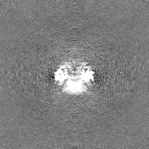
 emd_43271.png
 http://ftp.pdbj.org/pub/emdb/structures/EMD-43271
 Z (Sec.)
 Y (Row.)
 X (Col.)


 Sample components
 Processing
 Electron microscopy
 FIELD EMISSION GUN

