 Movie
Movie Controller


+ Open data

- Basic information
| Entry | 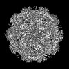 | ||||||||||||||||||
|---|---|---|---|---|---|---|---|---|---|---|---|---|---|---|---|---|---|---|---|
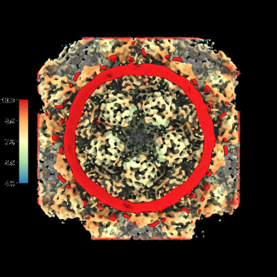
| Title | In situ STA of rotavirus enveloped DLP (penton) | ||||||||||||||||||
 Map data Map data | consensus average | ||||||||||||||||||
 Sample Sample |
| ||||||||||||||||||
| Biological species |  Rotavirus A Rotavirus A | ||||||||||||||||||
| Method | subtomogram averaging / cryo EM / Resolution: 10.2 Å | ||||||||||||||||||
 Authors Authors | Shah PNM / Stuart DI | ||||||||||||||||||
| Funding support |  United Kingdom, 5 items United Kingdom, 5 items
| ||||||||||||||||||
 Citation Citation | Journal: Cell Host Microbe / Year: 2023 Title: Characterization of the rotavirus assembly pathway in situ using cryoelectron tomography Authors: Shah PN / Gilchrist JB / Forsberg BO / Burt A / Howe A / Mosalaganti S / Wan W / Radecke J / Chaban Y / Sutton G / Stuart DI / Boyce M | ||||||||||||||||||
| History |
|
- Structure visualization
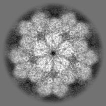
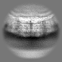
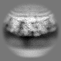
| Supplemental images |
|---|
- Downloads & links
-EMDB archive
| Map data | emd_16767.map.gz | 33 MB |  EMDB map data format EMDB map data format | |
|---|---|---|---|---|
| Header (meta data) | emd-16767-v30.xmlemd-16767.xml | 14.7 KB 14.7 KB | Display Display | EMDB header |
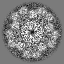
| Images |  emd_16767.png emd_16767.png | 182 KB | ||
| Others | emd_16767_half_map_1.map.gzemd_16767_half_map_2.map.gz | 17.7 MB 17.7 MB | ||
| Archive directory |  http://ftp.pdbj.org/pub/emdb/structures/EMD-16767ftp://ftp.pdbj.org/pub/emdb/structures/EMD-16767 http://ftp.pdbj.org/pub/emdb/structures/EMD-16767ftp://ftp.pdbj.org/pub/emdb/structures/EMD-16767 | HTTPS FTP |
-Links
| EMDB pages | EMDB (EBI/PDBe) / EMDataResource |
|---|
-Map
| File | Download / File: emd_16767.map.gz / Format: CCP4 / Size: 35.3 MB / Type: IMAGE STORED AS FLOATING POINT NUMBER (4 BYTES) | ||||||||||||||||||||||||||||||||||||
|---|---|---|---|---|---|---|---|---|---|---|---|---|---|---|---|---|---|---|---|---|---|---|---|---|---|---|---|---|---|---|---|---|---|---|---|---|---|
| Annotation | consensus average | ||||||||||||||||||||||||||||||||||||
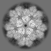
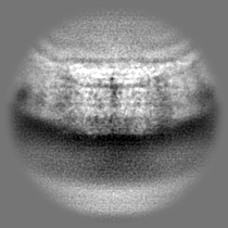
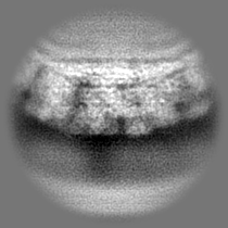
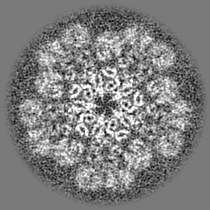
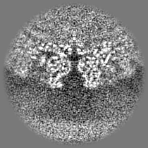
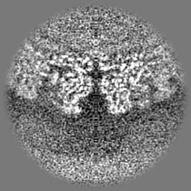
| Projections & slices | Image control
Images are generated by Spider. | ||||||||||||||||||||||||||||||||||||
| Voxel size | X=Y=Z: 1.97 Å | ||||||||||||||||||||||||||||||||||||
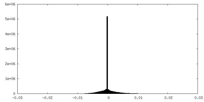
| Density |
| ||||||||||||||||||||||||||||||||||||
| Symmetry | Space group: 1 | ||||||||||||||||||||||||||||||||||||
| Details | EMDB XML:
|
 Z (Sec.)
 Y (Row.)
 X (Col.)


-Supplemental data
-Half map: half1
| File | emd_16767_half_map_1.map | ||||||||||||
|---|---|---|---|---|---|---|---|---|---|---|---|---|---|
| Annotation | half1 | ||||||||||||
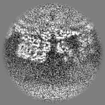
| Projections & Slices |
| ||||||||||||
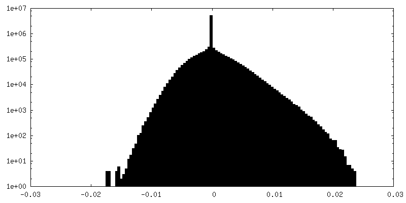
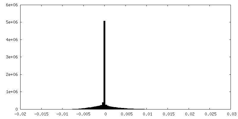
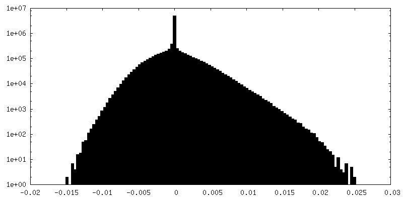
| Density Histograms |

-Half map: half2
| File | emd_16767_half_map_2.map | ||||||||||||
|---|---|---|---|---|---|---|---|---|---|---|---|---|---|
| Annotation | half2 | ||||||||||||
| Projections & Slices |
| ||||||||||||
| Density Histograms |
- Sample components
-Entire : Rotavirus A
| Entire | Name: Rotavirus A |
|---|---|
| Components |
|
-Supramolecule #1: Rotavirus A
| Supramolecule | Name: Rotavirus A / type: virus / ID: 1 / Parent: 0 / Macromolecule list: #1-#4 / NCBI-ID: 28875 / Sci species name: Rotavirus A / Virus type: VIRION / Virus isolate: STRAIN / Virus enveloped: No / Virus empty: No |
|---|
-Experimental details
-Structure determination
| Method | cryo EM |
|---|---|
 Processing Processing | subtomogram averaging |
| Aggregation state | cell |
-Sample preparation
| Buffer | pH: 7.5 |
|---|---|
| Grid | Model: Quantifoil R2/1 / Support film - Material: CARBON / Support film - topology: HOLEY |
| Vitrification | Cryogen name: ETHANE |
- Electron microscopy
| Microscope | TFS KRIOS |
|---|---|
| Image recording | Film or detector model: FEI FALCON IV (4k x 4k) / Detector mode: COUNTING / Average electron dose: 2.2 e/Å2 |
| Electron beam | Acceleration voltage: 300 kV / Electron source:  FIELD EMISSION GUN FIELD EMISSION GUN |
| Electron optics | Illumination mode: OTHER / Imaging mode: BRIGHT FIELD / Nominal defocus max: 3.5 µm / Nominal defocus min: 1.5 µm |
| Experimental equipment |  Model: Titan Krios / Image courtesy: FEI Company |
-Image processing
| Final reconstruction | Applied symmetry - Point group: C5 (5 fold cyclic) / Algorithm: BACK PROJECTION / Resolution.type: BY AUTHOR / Resolution: 10.2 Å / Resolution method: FSC 0.143 CUT-OFF / Software - Name: Warp (ver. 1.0.9) / Number subtomograms used: 138 |
|---|---|
| Extraction | Number tomograms: 85 / Number images used: 138 / Software - Name: crYOLO (ver. 1.8.2) |
| Final 3D classification | Software - Name: RELION (ver. 3.1.2) |
| Final angle assignment | Type: MAXIMUM LIKELIHOOD / Software - Name: RELION (ver. 3.1.2) |
