 ムービー
ムービー コントローラー
 構造ビューア
 EMN検索について


-検索条件
-検索結果
検索 (著者・登録者: mayer & a)の結果136件中、1から50件目までを表示しています

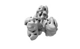
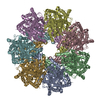
EMDB-43508: 
Structure of a bacterial gasdermin small oval pore assembly

EMDB-43509:
Structure of a bacterial gasdermin medium oval pore assembly

EMDB-43510:
Structure of a bacterial gasdermin large oval pore assembly

EMDB-43511:
Structure of a bacterial gasdermin double pore assembly

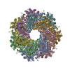
EMDB-43513:
Structure of a bacterial gasdermin slinky-like oligomer from a heterogeneous sample

EMDB-15411:
Single particle structure of Atg18-WT
PDB-8afx:
Single particle structure of Atg18-WT
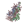
EMDB-18657:
PROTAC-mediated complex of KRAS with VHL/Elongin-B/Elongin-C/Cullin-2/Rbx1
EMDB-41983:
Structure of the phage immune evasion protein Gad1 bound to the Gabija GajAB complex
PDB-8u7i:
Structure of the phage immune evasion protein Gad1 bound to the Gabija GajAB complex

EMDB-15408:
Tube assembly of Atg18-PR72AA

EMDB-15410:
Tube assembly of Atg18-WT

EMDB-15412:
Subtomogram average of membrane-bound Atg18 oligomers
PDB-8afq:
Tube assembly of Atg18-PR72AA
PDB-8afw:
Tube assembly of Atg18-WT
PDB-8afy:
Subtomogram average of membrane-bound Atg18 oligomers

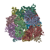
EMDB-17015:
Cryo-EM map of the focused refinement of the subfamily III haloalkane dehalogenase from Haloferax mediterranei dimer forming hexameric assembly.
PDB-8ooh:
Cryo-EM map of the focused refinement of the subfamily III haloalkane dehalogenase from Haloferax mediterranei dimer forming hexameric assembly.
EMDB-16998:
Cryo-EM structure of subfamily III haloalkane dehalogenase DhmeA from Haloferax mediterranei

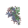
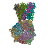
EMDB-27205:
Closed state of SARS-CoV-2 BA.2 variant spike protein

EMDB-27206:
One RBD-up state of SARS-CoV-2 BA.2 variant spike protein

EMDB-27207:
Middle state of SARS-CoV-2 BA.2 variant spike protein

PDB-8d55:
Closed state of SARS-CoV-2 BA.2 variant spike protein
PDB-8d56:
One RBD-up state of SARS-CoV-2 BA.2 variant spike protein
PDB-8d5a:
Middle state of SARS-CoV-2 BA.2 variant spike protein

EMDB-40570:
Structure of a bacterial gasdermin slinky-like oligomer

PDB-8sl0:
Structure of a bacterial gasdermin slinky-like oligomer
EMDB-29016:
Cryo-EM structure of SARS-CoV-2 postfusion spike in membrane
EMDB-29017:
Cryo-EM structure of SARS-CoV-2 postfusion spike in membrane

EMDB-29018:
Cryo-EM structure of SARS-CoV-2 postfusion spike in membrane

PDB-8fdw:
Cryo-EM structure of SARS-CoV-2 postfusion spike in membrane
EMDB-25419:
Previously uncharacterized rectangular bacteria in the dolphin mouth

EMDB-35208:
Cryo-EM structure of the polyphosphate polymerase VTC complex(Vtc4/Vtc3/Vtc1)

PDB-8i6v:
Cryo-EM structure of the polyphosphate polymerase VTC complex(Vtc4/Vtc3/Vtc1)
EMDB-29323:
Structure of RdrA from Escherichia coli RADAR defense system
EMDB-29324:
Map of RdrA from Escherichia coli RADAR defense system in single-split conformation
EMDB-29325:
Map of RdrA from Escherichia coli RADAR defense system in double-split conformation
EMDB-29326:
Structure of RdrA from Streptococcus suis RADAR defense system
EMDB-29327:
Structure of RdrB from Escherichia coli RADAR defense system
EMDB-29328:
Structure of RdrA-RdrB complex from Escherichia coli RADAR defense system
PDB-8fnt:
Structure of RdrA from Escherichia coli RADAR defense system
PDB-8fnu:
Structure of RdrA from Streptococcus suis RADAR defense system
PDB-8fnv:
Structure of RdrB from Escherichia coli RADAR defense system
PDB-8fnw:
Structure of RdrA-RdrB complex from Escherichia coli RADAR defense system

EMDB-27704:
Cryo-EM structure of insulin receptor (IR) bound with S597 peptide

EMDB-27705:
Cryo-EM structure of insulin receptor (IR) bound with S597 component 2

PDB-8dtl:
Cryo-EM structure of insulin receptor (IR) bound with S597 peptide
PDB-8dtm:
Cryo-EM structure of insulin receptor (IR) bound with S597 component 2
EMDB-31642:
Local construction of SARS-CoV-2 S protein RBD in complex with XG014 Fab

EMDB-25188:
Full-length insulin receptor bound with site 1 binding deficient mutant insulin (A-V3E)
ページ:
 wwPDBはEMDBデータモデルのバージョン3へ移行します
