Movie
Movie Controller

[English] 日本語
 Yorodumi
- EMDB-17015: Cryo-EM map of the focused refinement of the subfamily III haloal... -
+ Open data

- Basic information
| Entry |  | |||||||||
|---|---|---|---|---|---|---|---|---|---|---|
| Title | Cryo-EM map of the focused refinement of the subfamily III haloalkane dehalogenase from Haloferax mediterranei dimer forming hexameric assembly. | |||||||||
 Map data Map data | Cryo-EM map of the focused refinement of the subfamily III haloalkane dehalogenase from Haloferax mediterranei dimer forming hexameric assembly. | |||||||||
 Sample Sample |
| |||||||||
 Keywords Keywords | haloalkane dehalogenase / HYDROLASE | |||||||||
| Function / homology | haloalkane dehalogenase / haloalkane dehalogenase activity / : / Epoxide hydrolase-like / alpha/beta hydrolase fold / Alpha/beta hydrolase fold-1 / Alpha/Beta hydrolase fold / membrane / Alpha/beta fold hydrolase Function and homology information Function and homology information | |||||||||
| Biological species |  Haloferax mediterranei (archaea) Haloferax mediterranei (archaea) | |||||||||
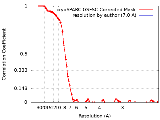
| Method | single particle reconstruction / cryo EM / Resolution: 7.0 Å | |||||||||
 Authors Authors | Polak M / Novacek J / Chmelova K / Marek M | |||||||||
| Funding support |  Czech Republic, 1 items Czech Republic, 1 items
| |||||||||
 Citation Citation | Journal: Protein Sci / Year: 2023 Title: Multimeric structure of a subfamily III haloalkane dehalogenase-like enzyme solved by combination of cryo-EM and x-ray crystallography. Authors: Klaudia Chmelova / Tadeja Gao / Martin Polak / Andrea Schenkmayerova / Tristan I Croll / Tanvir R Shaikh / Jana Skarupova / Radka Chaloupkova / Kay Diederichs / Randy J Read / Jiri Damborsky ...Authors: Klaudia Chmelova / Tadeja Gao / Martin Polak / Andrea Schenkmayerova / Tristan I Croll / Tanvir R Shaikh / Jana Skarupova / Radka Chaloupkova / Kay Diederichs / Randy J Read / Jiri Damborsky / Jiri Novacek / Martin Marek /   Abstract: Haloalkane dehalogenase (HLD) enzymes employ an S 2 nucleophilic substitution mechanism to erase halogen substituents in diverse organohalogen compounds. Subfamily I and II HLDs are well- ...Haloalkane dehalogenase (HLD) enzymes employ an S 2 nucleophilic substitution mechanism to erase halogen substituents in diverse organohalogen compounds. Subfamily I and II HLDs are well-characterized enzymes, but the mode and purpose of multimerization of subfamily III HLDs are unknown. Here we probe the structural organization of DhmeA, a subfamily III HLD-like enzyme from the archaeon Haloferax mediterranei, by combining cryo-electron microscopy (cryo-EM) and x-ray crystallography. We show that full-length wild-type DhmeA forms diverse quaternary structures, ranging from small oligomers to large supramolecular ring-like assemblies of various sizes and symmetries. We optimized sample preparation steps, enabling three-dimensional reconstructions of an oligomeric species by single-particle cryo-EM. Moreover, we engineered a crystallizable mutant (DhmeA ) that provided diffraction-quality crystals. The 3.3 Å crystal structure reveals that DhmeA forms a ring-like 20-mer structure with outer and inner diameter of ~200 and ~80 Å, respectively. An enzyme homodimer represents a basic repeating building unit of the crystallographic ring. Three assembly interfaces (dimerization, tetramerization, and multimerization) were identified to form the supramolecular ring that displays a negatively charged exterior, while its interior part harboring catalytic sites is positively charged. Localization and exposure of catalytic machineries suggest a possible processing of large negatively charged macromolecular substrates. | |||||||||
| History |
|
- Structure visualization
| Supplemental images |
|---|

- Downloads & links
-EMDB archive
| Map data | emd_17015.map.gz | 30.3 MB | EMDB map data format | |
|---|---|---|---|---|
| Header (meta data) | emd-17015-v30.xmlemd-17015.xml | 15.4 KB 15.4 KB | Display Display | EMDB header |
| FSC (resolution estimation) | emd_17015_fsc.xml | 8.5 KB | Display | FSC data file |
| Images | 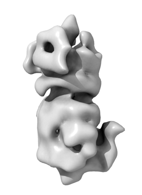 emd_17015.png emd_17015.png | 38.8 KB | ||
| Filedesc metadata | emd-17015.cif.gz | 5.5 KB | ||
| Others | emd_17015_half_map_1.map.gzemd_17015_half_map_2.map.gz | 59.3 MB 59.3 MB | ||
| Archive directory |  http://ftp.pdbj.org/pub/emdb/structures/EMD-17015ftp://ftp.pdbj.org/pub/emdb/structures/EMD-17015 http://ftp.pdbj.org/pub/emdb/structures/EMD-17015ftp://ftp.pdbj.org/pub/emdb/structures/EMD-17015 | HTTPS FTP |
-Related structure data
| Related structure data | 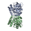 8oohMC 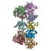 8ckpC M: atomic model generated by this map C: citing same article ( |
|---|---|
| Similar structure data |

-Links
| EMDB pages | EMDB (EBI/PDBe) / EMDataResource |
|---|---|
| Related items in Molecule of the Month |


-Map
| File | Download / File: emd_17015.map.gz / Format: CCP4 / Size: 64 MB / Type: IMAGE STORED AS FLOATING POINT NUMBER (4 BYTES) | ||||||||||||||||||||||||||||||||||||
|---|---|---|---|---|---|---|---|---|---|---|---|---|---|---|---|---|---|---|---|---|---|---|---|---|---|---|---|---|---|---|---|---|---|---|---|---|---|
| Annotation | Cryo-EM map of the focused refinement of the subfamily III haloalkane dehalogenase from Haloferax mediterranei dimer forming hexameric assembly. | ||||||||||||||||||||||||||||||||||||
| Projections & slices | Image control
Images are generated by Spider. | ||||||||||||||||||||||||||||||||||||
| Voxel size | X=Y=Z: 1.061 Å | ||||||||||||||||||||||||||||||||||||
| Density |
| ||||||||||||||||||||||||||||||||||||
| Symmetry | Space group: 1 | ||||||||||||||||||||||||||||||||||||
| Details | EMDB XML:
|
 Z (Sec.)
 Y (Row.)
 X (Col.)


-Supplemental data
-Half map: Second half map.
| File | emd_17015_half_map_1.map | ||||||||||||
|---|---|---|---|---|---|---|---|---|---|---|---|---|---|
| Annotation | Second half map. | ||||||||||||
| Projections & Slices |
| ||||||||||||
| Density Histograms |


-Half map: First half map.
| File | emd_17015_half_map_2.map | ||||||||||||
|---|---|---|---|---|---|---|---|---|---|---|---|---|---|
| Annotation | First half map. | ||||||||||||
| Projections & Slices |
| ||||||||||||
| Density Histograms |


- Sample components
-Entire : The subfamily III haloalkane dehalogenase DhmeA from Haloferax me...
| Entire | Name: The subfamily III haloalkane dehalogenase DhmeA from Haloferax mediterranei |
|---|---|
| Components |
|
-Supramolecule #1: The subfamily III haloalkane dehalogenase DhmeA from Haloferax me...
| Supramolecule | Name: The subfamily III haloalkane dehalogenase DhmeA from Haloferax mediterranei type: complex / ID: 1 / Parent: 0 / Macromolecule list: all |
|---|---|
| Source (natural) | Organism: Haloferax mediterranei (archaea) |
-Macromolecule #1: Alpha/beta fold hydrolase
| Macromolecule | Name: Alpha/beta fold hydrolase / type: protein_or_peptide / ID: 1 / Number of copies: 2 / Enantiomer: LEVO |
|---|---|
| Source (natural) | Organism: Haloferax mediterranei (archaea) |
| Molecular weight | Theoretical: 35.711797 KDa |
| Recombinant expression | Organism:  |
| Sequence | String: MSSASSNARD EVIAAIHEEA DWVDRTVYPF ESRCIGLSSG AVHYIDEGPD DGGRETLLML HGNPTWSFLY RHLVRDLRDE YRCVALDYL GFGLSERPTD FSYRPEDHAD VVEEFIDELG LEDVVLVGHD WGGPIGFSYA IDHPENVGGL VVMNTWMWPV S DDKHFSRF ...String: MSSASSNARD EVIAAIHEEA DWVDRTVYPF ESRCIGLSSG AVHYIDEGPD DGGRETLLML HGNPTWSFLY RHLVRDLRDE YRCVALDYL GFGLSERPTD FSYRPEDHAD VVEEFIDELG LEDVVLVGHD WGGPIGFSYA IDHPENVGGL VVMNTWMWPV S DDKHFSRF SKLLGGRIGR ELCERYDLFT RVIMPMGFAD RSRFTESARE QYRAANRGDR TGTGIFPQAI LGSRAWLSSL WE QRDNIAD IPARIIWGME DSAFRPAELR TFEALFEDSS TVRLYGVGHY VPEEFGSDLV PLVREFLEEV LEVLFQ UniProtKB: Alpha/beta fold hydrolase |
-Experimental details
-Structure determination
| Method | cryo EM |
|---|---|
 Processing Processing | single particle reconstruction |
| Aggregation state | particle |
-Sample preparation
| Concentration | 0.035 mg/mL |
|---|---|
| Buffer | pH: 8 / Component - Concentration: 1.0 M / Component - Formula: NaCl / Component - Name: Sodium Chloride / Details: 1 M NaCl, 10 mM Tris-HCl |
| Vitrification | Cryogen name: ETHANE |
| Details | This sample was purified by gel filtration. |
- Electron microscopy
| Microscope | FEI TITAN KRIOS |
|---|---|
| Image recording | #0 - Image recording ID: 1 / #0 - Film or detector model: FEI FALCON II (4k x 4k) / #0 - Average electron dose: 48.0 e/Å2 / #1 - Image recording ID: 2 / #1 - Film or detector model: GATAN K2 QUANTUM (4k x 4k) / #1 - Average electron dose: 73.0 e/Å2 |
| Electron beam | Acceleration voltage: 300 kV / Electron source:  FIELD EMISSION GUN FIELD EMISSION GUN |
| Electron optics | Illumination mode: FLOOD BEAM / Imaging mode: BRIGHT FIELD / Nominal defocus max: 2.2 µm / Nominal defocus min: 1.0 µm |
| Experimental equipment |  Model: Titan Krios / Image courtesy: FEI Company |