[English] 日本語
Yorodumi
- PDB-8ckp: X-ray structure of the crystallization-prone form of subfamily II... -

+
Open data


ID or keywords:

Loading...

-
Basic information

Entry
Database: PDB / ID: 8ckp
TitleX-ray structure of the crystallization-prone form of subfamily III haloalkane dehalogenase DhmeA from Haloferax mediterranei
ComponentsAlpha/beta fold hydrolase
KeywordsUNKNOWN FUNCTION / haloalkane dehalogenase-like / hydrolase
Function / homologyhaloalkane dehalogenase / haloalkane dehalogenase activity / acyltransferase activity / Epoxide hydrolase-like / alpha/beta hydrolase fold / Alpha/beta hydrolase fold-1 / Alpha/Beta hydrolase fold / Alpha/beta fold hydrolase
Function and homology information
Biological speciesHaloferax mediterranei (archaea)
MethodX-RAY DIFFRACTION / SYNCHROTRON / MOLECULAR REPLACEMENT / Resolution: 3.31 Å
AuthorsMarek, M. / Chmelova, K. / Schenkmayerova, A. / Croll, T. / Read, R.J. / Diederichs, K.
Funding support Czech Republic, 1items
OrganizationGrant numberCountry
Czech Science FoundationGA22-09853S Czech Republic
Citation
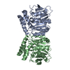
Journal: Protein Sci / Year: 2023
Title: Multimeric structure of a subfamily III haloalkane dehalogenase-like enzyme solved by combination of cryo-EM and x-ray crystallography.
Authors: Klaudia Chmelova / Tadeja Gao / Martin Polak / Andrea Schenkmayerova / Tristan I Croll / Tanvir R Shaikh / Jana Skarupova / Radka Chaloupkova / Kay Diederichs / Randy J Read / Jiri Damborsky ...Authors: Klaudia Chmelova / Tadeja Gao / Martin Polak / Andrea Schenkmayerova / Tristan I Croll / Tanvir R Shaikh / Jana Skarupova / Radka Chaloupkova / Kay Diederichs / Randy J Read / Jiri Damborsky / Jiri Novacek / Martin Marek /
Abstract: Haloalkane dehalogenase (HLD) enzymes employ an S 2 nucleophilic substitution mechanism to erase halogen substituents in diverse organohalogen compounds. Subfamily I and II HLDs are well- ...Haloalkane dehalogenase (HLD) enzymes employ an S 2 nucleophilic substitution mechanism to erase halogen substituents in diverse organohalogen compounds. Subfamily I and II HLDs are well-characterized enzymes, but the mode and purpose of multimerization of subfamily III HLDs are unknown. Here we probe the structural organization of DhmeA, a subfamily III HLD-like enzyme from the archaeon Haloferax mediterranei, by combining cryo-electron microscopy (cryo-EM) and x-ray crystallography. We show that full-length wild-type DhmeA forms diverse quaternary structures, ranging from small oligomers to large supramolecular ring-like assemblies of various sizes and symmetries. We optimized sample preparation steps, enabling three-dimensional reconstructions of an oligomeric species by single-particle cryo-EM. Moreover, we engineered a crystallizable mutant (DhmeA ) that provided diffraction-quality crystals. The 3.3 Å crystal structure reveals that DhmeA forms a ring-like 20-mer structure with outer and inner diameter of ~200 and ~80 Å, respectively. An enzyme homodimer represents a basic repeating building unit of the crystallographic ring. Three assembly interfaces (dimerization, tetramerization, and multimerization) were identified to form the supramolecular ring that displays a negatively charged exterior, while its interior part harboring catalytic sites is positively charged. Localization and exposure of catalytic machineries suggest a possible processing of large negatively charged macromolecular substrates.
History
DepositionFeb 16, 2023Deposition site: PDBE / Processing site: PDBE
Revision 1.0Aug 30, 2023Provider: repository / Type: Initial release
Revision 1.1Oct 11, 2023Group: Database references / Category: citation / citation_author
Item: _citation.journal_volume / _citation.title / _citation_author.identifier_ORCID

-
Structure visualization

Structure viewerMolecule:
MolmilJmol/JSmol

Downloads & links

-
Assembly

Deposited unit
D: Alpha/beta fold hydrolase
F: Alpha/beta fold hydrolase
G: Alpha/beta fold hydrolase
H: Alpha/beta fold hydrolase
I: Alpha/beta fold hydrolase
J: Alpha/beta fold hydrolase
A: Alpha/beta fold hydrolase
B: Alpha/beta fold hydrolase
C: Alpha/beta fold hydrolase
E: Alpha/beta fold hydrolase
hetero molecules


Theoretical massNumber of molelcules
Total (without water)357,00311
Polymers356,96710
Non-polymers351
Water181
1


  • Idetical with deposited unit
  • defined by author
  • Evidence: gel filtration
TypeNameSymmetry operationNumber
identity operation1_555x,y,z1
Buried area19260 Å2
ΔGint-51 kcal/mol
Surface area107700 Å2
Unit cell
Length a, b, c (Å)172.760, 289.900, 168.310
Angle α, β, γ (deg.)90.000, 90.000, 90.000
Int Tables number20
Space group name H-MC2221
Space group name HallC2c2
Symmetry operation#1: x,y,z
#2: x,-y,-z
#3: -x,y,-z+1/2
#4: -x,-y,z+1/2
#5: x+1/2,y+1/2,z
#6: x+1/2,-y+1/2,-z
#7: -x+1/2,y+1/2,-z+1/2
#8: -x+1/2,-y+1/2,z+1/2

-
Components

#1: Protein
Alpha/beta fold hydrolase


Mass: 35696.707 Da / Num. of mol.: 10
Source method: isolated from a genetically manipulated source
Source: (gene. exp.) Haloferax mediterranei (archaea) / Gene: mhpC / Production host: Escherichia coli (E. coli) / References: UniProt: I3R766
#2: Chemical ChemComp-CL / CHLORIDE ION / Chloride


Mass: 35.453 Da / Num. of mol.: 1 / Source method: obtained synthetically / Formula: Cl / Feature type: SUBJECT OF INVESTIGATION
#3: Water ChemComp-HOH / water / Water


Mass: 18.015 Da / Num. of mol.: 1 / Source method: isolated from a natural source / Formula: H2O
Has ligand of interestY

-
Experimental details

-
Experiment

ExperimentMethod: X-RAY DIFFRACTION / Number of used crystals: 1

-
Sample preparation

CrystalDensity Matthews: 2.95 Å3/Da / Density % sol: 58.33 %
Crystal growTemperature: 293.15 K / Method: vapor diffusion, sitting drop / pH: 6.5 / Details: PEG 4000, MES/Imidazole buffer system, amino acids

-
Data collection

DiffractionMean temperature: 100 K / Serial crystal experiment: N
Diffraction sourceSource: SYNCHROTRON / Site: SLS / Beamline: X06DA / Wavelength: 0.9999 Å
DetectorType: DECTRIS PILATUS 2M-F / Detector: PIXEL / Date: May 12, 2019
RadiationProtocol: SINGLE WAVELENGTH / Monochromatic (M) / Laue (L): M / Scattering type: x-ray
Radiation wavelengthWavelength: 0.9999 Å / Relative weight: 1
ReflectionResolution: 3.306→49.47 Å / Num. obs: 63328 / % possible obs: 99.55 % / Redundancy: 70.3 % / Biso Wilson estimate: 151.61 Å2 / CC1/2: 0.999 / Net I/σ(I): 10.76
Reflection shellResolution: 3.306→3.424 Å / Num. unique obs: 6044 / CC1/2: 0.241

-
Processing

Software
NameVersionClassification
XDSdata scaling
XDSdata reduction
PHASERphasing
PHENIX1.20.1-4487refinement
RefinementMethod to determine structure: MOLECULAR REPLACEMENT / Resolution: 3.31→49.47 Å / SU ML: 0.6973 / Cross valid method: FREE R-VALUE / σ(F): 1.33 / Phase error: 36.1909
Stereochemistry target values: GeoStd + Monomer Library + CDL v1.2
RfactorNum. reflection% reflection
Rfree0.2748 3153 5 %
Rwork0.2358 59920 -
obs0.2379 63073 99.59 %
Solvent computationShrinkage radii: 0.9 Å / VDW probe radii: 1.11 Å / Solvent model: FLAT BULK SOLVENT MODEL
Displacement parametersBiso mean: 155.02 Å2
Refinement stepCycle: LAST / Resolution: 3.31→49.47 Å
ProteinNucleic acidLigandSolventTotal
Num. atoms23882 0 1 1 23884
Refine LS restraints
Refine-IDTypeDev idealNumber
X-RAY DIFFRACTIONf_bond_d0.002524550
X-RAY DIFFRACTIONf_angle_d0.691633342
X-RAY DIFFRACTIONf_chiral_restr0.04543439
X-RAY DIFFRACTIONf_plane_restr0.01194420
X-RAY DIFFRACTIONf_dihedral_angle_d13.28838833
LS refinement shell
Resolution (Å)Rfactor RfreeNum. reflection RfreeRfactor RworkNum. reflection RworkRefine-ID% reflection obs (%)
3.31-3.360.50281240.54412394X-RAY DIFFRACTION93.26
3.36-3.410.53271360.46622569X-RAY DIFFRACTION99.41
3.41-3.460.4491360.41622590X-RAY DIFFRACTION99.96
3.46-3.520.43791370.41082602X-RAY DIFFRACTION99.96
3.52-3.590.43961360.35582585X-RAY DIFFRACTION99.96
3.59-3.660.38041370.34712602X-RAY DIFFRACTION99.93
3.66-3.730.45231360.35632583X-RAY DIFFRACTION99.78
3.73-3.810.34621350.292556X-RAY DIFFRACTION99.85
3.81-3.90.36371370.30552611X-RAY DIFFRACTION99.96
3.9-40.38261360.31592579X-RAY DIFFRACTION99.52
4-4.110.36311380.26322620X-RAY DIFFRACTION99.86
4.11-4.230.30911360.24552586X-RAY DIFFRACTION99.96
4.23-4.360.26211360.22532583X-RAY DIFFRACTION99.96
4.36-4.520.29121370.22312604X-RAY DIFFRACTION100
4.52-4.70.28821380.22222616X-RAY DIFFRACTION99.93
4.7-4.910.28181370.22292604X-RAY DIFFRACTION99.93
4.91-5.170.23851390.19482635X-RAY DIFFRACTION99.93
5.17-5.50.24891370.22182616X-RAY DIFFRACTION100
5.5-5.920.29431390.23382640X-RAY DIFFRACTION100
5.92-6.510.24011400.21982647X-RAY DIFFRACTION99.96
6.52-7.450.28051390.21872641X-RAY DIFFRACTION99.96
7.45-9.380.21161410.1792690X-RAY DIFFRACTION100
9.38-49.470.21461460.21192767X-RAY DIFFRACTION99.49

+
About Yorodumi

-
News

-
Feb 9, 2022. New format data for meta-information of EMDB entries

New format data for meta-information of EMDB entries

  • Version 3 of the EMDB header file is now the official format.
  • The previous official version 1.9 will be removed from the archive.

Related info.:EMDB header

External links:wwPDB to switch to version 3 of the EMDB data model

-
Aug 12, 2020. Covid-19 info

Covid-19 info

URL: https://pdbj.org/emnavi/covid19.php

New page: Covid-19 featured information page in EM Navigator.

Related info.:Covid-19 info / Mar 5, 2020. Novel coronavirus structure data

+
Mar 5, 2020. Novel coronavirus structure data

Novel coronavirus structure data

Related info.:Yorodumi Speices / Aug 12, 2020. Covid-19 info

External links:COVID-19 featured content - PDBj / Molecule of the Month (242):Coronavirus Proteases

+
Jan 31, 2019. EMDB accession codes are about to change! (news from PDBe EMDB page)

EMDB accession codes are about to change! (news from PDBe EMDB page)

  • The allocation of 4 digits for EMDB accession codes will soon come to an end. Whilst these codes will remain in use, new EMDB accession codes will include an additional digit and will expand incrementally as the available range of codes is exhausted. The current 4-digit format prefixed with “EMD-” (i.e. EMD-XXXX) will advance to a 5-digit format (i.e. EMD-XXXXX), and so on. It is currently estimated that the 4-digit codes will be depleted around Spring 2019, at which point the 5-digit format will come into force.
  • The EM Navigator/Yorodumi systems omit the EMD- prefix.

Related info.:Q: What is EMD? / ID/Accession-code notation in Yorodumi/EM Navigator

External links:EMDB Accession Codes are Changing Soon! / Contact to PDBj

+
Jul 12, 2017. Major update of PDB

Major update of PDB

  • wwPDB released updated PDB data conforming to the new PDBx/mmCIF dictionary.
  • This is a major update changing the version number from 4 to 5, and with Remediation, in which all the entries are updated.
  • In this update, many items about electron microscopy experimental information are reorganized (e.g. em_software).
  • Now, EM Navigator and Yorodumi are based on the updated data.

External links:wwPDB Remediation / Enriched Model Files Conforming to OneDep Data Standards Now Available in the PDB FTP Archive

-
Yorodumi

Thousand views of thousand structures

  • Yorodumi is a browser for structure data from EMDB, PDB, SASBDB, etc.
  • This page is also the successor to EM Navigator detail page, and also detail information page/front-end page for Omokage search.
  • The word "yorodu" (or yorozu) is an old Japanese word meaning "ten thousand". "mi" (miru) is to see.

Related info.:EMDB / PDB / SASBDB / Comparison of 3 databanks / Yorodumi Search / Aug 31, 2016. New EM Navigator & Yorodumi / Yorodumi Papers / Jmol/JSmol / Function and homology information / Changes in new EM Navigator and Yorodumi

Read more