 ムービー
ムービー コントローラー
 構造ビューア
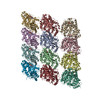
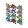
 EMN検索について


-検索条件
-検索結果
検索 (著者・登録者: massimi & a)の結果全30件を表示しています
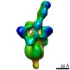
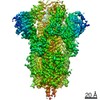
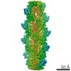
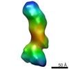
EMDB-23523: 
SARS-2 CoV 6P Mut7 in complex with Fab J08
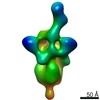
EMDB-23524:
SARS-2 CoV 6P Mut7 in complex with Fab F05
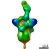
EMDB-23525:
SARS-2 CoV 6P Mut 7 in complex with fragment antigen binding (fab) I14

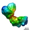
EMDB-12142:
ASCT2 in the presence of the inhibitor Lc-BPE in the outward-open conformation.

EMDB-12143:
ASCT2 in the presence of the inhibitor ERA-21 in the outward-open conformation.
PDB-7bcq:
ASCT2 in the presence of the inhibitor Lc-BPE (position "up") in the outward-open conformation.
PDB-7bcs:
ASCT2 in the presence of the inhibitor Lc-BPE (position "down") in the outward-open conformation.
PDB-7bct:
ASCT2 in the presence of the inhibitor ERA-21 in the outward-open conformation.
EMDB-22078:
Characterization of the SARS-CoV-2 S Protein: Biophysical, Biochemical, Structural, and Antigenic Analysis

PDB-6x6p:
Characterization of the SARS-CoV-2 S Protein: Biophysical, Biochemical, Structural, and Antigenic Analysis
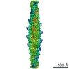
EMDB-0612:
CryoEM Density of Acetylated Microtubules

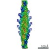
EMDB-0613:
Deacetylated Microtubules

EMDB-0614:
Deacetylated Microtubules
EMDB-0615:
Acetylated Microtubules
PDB-6o2q:
Acetylated Microtubules
PDB-6o2r:
Deacetylated Microtubules
PDB-6o2s:
Deacetylated Microtubules

PDB-6o2t:
Acetylated Microtubules
EMDB-7831:
Cryo-EM structure of FLNaABD E254K bound to phalloidin-stabilized F-actin
EMDB-7832:
FLNaABD-Q170P bound to phalloidin-stabilized F-actin

EMDB-7833:
FLNaABD-WT bound to phalloidin-stabilized F-actin
EMDB-8918:
Cryo-EM structure of FLNa ABD-E254K bound to phalloidin-stabilized F-actin, collected on a FEI Tecnai F20 microscope

PDB-6d8c:
Cryo-EM structure of FLNaABD E254K bound to phalloidin-stabilized F-actin
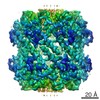
EMDB-7950:
cryo-EM density map of ClpP from Staphylococcus aureus (Wild Type)

EMDB-7951:
cryo-EM density map of ClpP from Staphylococcus aureus with bound Acyldepsipeptide (V7A mutant)

EMDB-7952:
Caseinolytic protease (ClpP) from Staphylococcus aureus mutant - V7A

PDB-6dkf:
Caseinolytic protease (ClpP) from Staphylococcus aureus mutant - V7A
EMDB-3391:
Negative-stain electron microscopy structure of human cytomegalovirus gHgLgO trimer
EMDB-2631:
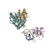
The Cryo-EM structure of the palindromic DNA-bound USP/EcR nuclear receptor reveals an asymmetric organization with allosteric domain positioning
PDB-4umm:
The Cryo-EM structure of the palindromic DNA-bound USP-EcR nuclear receptor reveals an asymmetric organization with allosteric domain positioning
 wwPDBはEMDBデータモデルのバージョン3へ移行します
