Movie
Movie Controller
 Structure viewers
 About EMN search



-Search query
-Search result
Showing 1 - 50 of 191 items for (author: lv & mj)

EMDB-53847: 
Cryo-EM structure of human ATP citrate lyase in complex with inhibitor EVT0185-CoA
Method: single particle / : Verstraete K, Verschueren K, Savvides SN, Steinberg GR

EMDB-72725:
Cryo-EM structure of ternary complex BCL6-CRBN-DDB1 with BMS-986458 (local refined), a potent and selective BCL6 ligand directed degrader (LDD)
Method: single particle / : Zhu J, Fang W, Pagarigan B

PDB-9ya9:
Cryo-EM structure of ternary complex BCL6-CRBN-DDB1 with BMS-986458 (local refined), a potent and selective BCL6 ligand directed degrader (LDD)
Method: single particle / : Zhu J, Fang W, Pagarigan B

EMDB-52253:
Influenza Neuraminidase in complex with N-Acyl Oseltamivir inhibitor
Method: single particle / : Moran E, Davies G

EMDB-52254:
Influenza Neuraminidase in complex with N-Acyl Oseltamivir inhibitor
Method: single particle / : Moran E, Davies G

EMDB-52255:
Influenza Neuraminidase in complex with N-Acyl Oseltamivir inhibitor
Method: single particle / : Moran E, Davies G

EMDB-49708:
cryo-EM structure of broad betacoronavirus binding antibody 1871 in complex with OC43 S2 subunit
Method: single particle / : Muthuraman K, Jackman MJ, Julien JP

EMDB-54375:
Tomogram showing an NA membrane in an A549wt cell infected with WSNdeltaHA at 16 hpi.
Method: electron tomography / : Wachsmuth-Melm M, Chlanda P

EMDB-51740:
Subtomogram average of nuclear helical M1 assemblies
Method: subtomogram averaging / : Wachsmuth-Melm M, Chlanda P

EMDB-51741:
Subtomogram average of zippered influenza A virus neuraminidase
Method: subtomogram averaging / : Wachsmuth-Melm M, Chlanda P

EMDB-51742:
Subtomogram average of influenza vRNPs in-situ.
Method: subtomogram averaging / : Wachsmuth-Melm M, Chlanda P

EMDB-51790:
Subtomogram average of intracellular influenza A virus hemagglutinin (subtype H1)
Method: subtomogram averaging / : Wachsmuth-Melm M, Chlanda P

EMDB-51811:
Tomogram showing M1 cylinders in a VeroE6 cell transfected with M1 (HK68) 24 hpt [figure 3].
Method: electron tomography / : Wachsmuth-Melm M, Chlanda P

EMDB-70018:
BG505-DS SOSIP in complex with 007 bNAb Fabs - Class 0 (unbound)
Method: single particle / : DeLaitsch AT, Bjorkman PJ

EMDB-70019:
BG505-DS SOSIP in complex with 007 bNAb Fabs - Class 1 (1 Fab bound)
Method: single particle / : DeLaitsch AT, Bjorkman PJ

EMDB-70020:
BG505-DS SOSIP in complex with 007 bNAb Fabs - Class 2 (2 Fabs bound)
Method: single particle / : DeLaitsch AT, Bjorkman PJ

EMDB-70021:
BG505-DS SOSIP in complex with 007 bNAb Fabs - Class 3 (3 Fabs bound)
Method: single particle / : DeLaitsch AT, Bjorkman PJ

EMDB-70022:
BG505 SOSIP in complex with 007 bNAb IgG1 - trimer-dimer class
Method: single particle / : DeLaitsch AT, Bjorkman PJ

EMDB-52201:
Cryo-EM structure of CDK2-cyclin A bound to a GMNC peptide
Method: single particle / : de Martin Garrido N, Ord M, Cushing VI, Greber BJ, Pryciak PM, Davey NE

EMDB-52204:
Cryo-EM structure of CDK2-cyclin A bound to a SAMHD1 peptide
Method: single particle / : de Martin Garrido N, Ord M, Cushing VI, Greber BJ, Pryciak PM, Davey NE

EMDB-52208:
Cryo-EM structure of CDK2-cyclin A bound to a SCAPER peptide
Method: single particle / : de Martin Garrido N, Ord M, Cushing VI, Greber BJ, Pryciak PM, Davey NE

EMDB-52621:
Toxoplasma gondii cytochrome bc1 complex from the respiratory supercomplex III2-IV inhibited by atovaquone and ELQ-300
Method: single particle / : Maclean A, Muhleip A

EMDB-51939:
Cryo-EM structure of the atovaquone-inhibited Complex III from the Chlorocebus sabaeus respirasome
Method: single particle / : Maclean A, Muhleip A

EMDB-51157:
Cryo-EM structure of the Toxoplasma gondii respiratory chain complex III inhibited by ELQ-300
Method: single particle / : MacLean A, Muhleip A
EMDB-48283:
61-12A01 Fab in complex with HIV-1 GT1.1 v4.1 SOSIP Env trimer and RM20A3 Fab
Method: single particle / : Phulera S, Ozorowski G, Ward AB
EMDB-48286:
206-3G08 Fab in complex with HIV-1 GT1.1 v4.1 SOSIP Env trimer and RM20A3 Fab
Method: single particle / : Phulera S, Ozorowski G, Ward AB
EMDB-48287:
206-9C09 Fab in complex with HIV-1 GT1.1 v4.1 SOSIP Env trimer and RM20A3 Fab
Method: single particle / : Phulera S, Ozorowski G, Ward AB
EMDB-48290:
273-4D01 Fab in complex with HIV-1 BG505 SOSIP Env trimer and RM20A3 Fab
Method: single particle / : Phulera S, Ozorowski G, Ward AB
EMDB-48291:
253-7A03 Fab in complex with HIV-1 BG505 SOSIP Env trimer and RM20A3 Fab
Method: single particle / : Phulera S, Ozorowski G, Ward AB
EMDB-70490:
BG505 GT1.1 SOSIP in complex with gp41-base epitope polyclonal antibodies isolated from a participant in the IAVI C101 clinical trial
Method: single particle / : Ozorowski G, Ward AB
EMDB-70491:
BG505 GT1.1 SOSIP in complex with V1V2V3 epitope polyclonal antibodies isolated from a participant in the IAVI C101 clinical trial
Method: single particle / : Ozorowski G, Ward AB
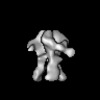
EMDB-70492:
BG505 GT1.1 SOSIP in complex with C3V5 epitope polyclonal antibodies isolated from a participant in the IAVI C101 clinical trial
Method: single particle / : Ozorowski G, Ward AB
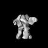
EMDB-70493:
BG505 GT1.1 SOSIP in complex with CD4bs epitope polyclonal antibodies isolated from a participant in the IAVI C101 clinical trial
Method: single particle / : Ozorowski G, Ward AB
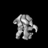
EMDB-70494:
BG505 GT1.1 SOSIP in complex with gp41 glycan hole epitope polyclonal antibodies isolated from a participant in the IAVI C101 clinical trial
Method: single particle / : Ozorowski G, Ward AB
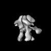
EMDB-70495:
BG505 GT1.1 SOSIP in complex with gp41 fusion peptide epitope polyclonal antibodies isolated from a participant in the IAVI C101 clinical trial
Method: single particle / : Ozorowski G, Ward AB

EMDB-50067:
Tomogram showing an HA remodelled membrane in an A549wt cell infected with PR8 at 16 hpi [figure 1].
Method: electron tomography / : Wachsmuth-Melm M, Chlanda P

EMDB-50070:
Tomogram showing an HA remodelled membrane in an A549wt cell infected with HK68 at 16 hpi [figure 1].
Method: electron tomography / : Wachsmuth-Melm M, Chlanda P

EMDB-50078:
Tomogram showing an HA remodelled membrane in an A549wt cell infected with HK68 at 16 hpi [figure 1].
Method: electron tomography / : Wachsmuth-Melm M, Chlanda P

EMDB-50079:
Tomogram showing an HA remodelled membrane in an A549wt cell infected with HK68 at 16 hpi [figure 1].
Method: electron tomography / : Wachsmuth-Melm M, Chlanda P

EMDB-50080:
Tomogram showing an HA remodelled membrane in an A549wt cell infected with PR8 at 16 hpi [figure 1].
Method: electron tomography / : Wachsmuth-Melm M, Chlanda P

EMDB-50081:
Tomogram showing an HA remodelled membrane in an A549-Rab11wt cell infected with PR8 at 16 hpi [figure 2].
Method: electron tomography / : Wachsmuth-Melm M, Chlanda P

EMDB-50082:
A549-Rab11dn cell infected with PR8 virus at 16 hpi.
Method: electron tomography / : Wachsmuth-Melm M, Chlanda P

EMDB-50083:
A549wt cell infected with PR8 virus at 16 hpi.
Method: electron tomography / : Wachsmuth-Melm M, Chlanda P

EMDB-50084:
A549wt cell infected with PR8 virus at 8 hpi.
Method: electron tomography / : Wachsmuth-Melm M, Chlanda P

EMDB-50085:
A549wt cell infected with PR8 virus at 16 hpi.
Method: electron tomography / : Wachsmuth-Melm M, Chlanda P

EMDB-50086:
A549wt cell infected with PR8 virus at 16 hpi.
Method: electron tomography / : Wachsmuth-Melm M, Chlanda P

EMDB-50087:
A549wt cell infected with PR8 virus at 16 hpi.
Method: electron tomography / : Wachsmuth-Melm M, Chlanda P

EMDB-50088:
A549wt cell infected with HK68 virus at 16 hpi.
Method: electron tomography / : Wachsmuth-Melm M, Chlanda P

EMDB-44403:
Cryo-EM of Hyper2 tube, ~27 nm diameter
Method: helical / : Sonani RR, Miller JG, Conticello V, Egelman EH

EMDB-44404:
Cryo-EM of Hyper2 tube, ~24 nm diameter
Method: helical / : Sonani RR, Miller JG, Conticello V, Egelman EH
Pages:
 wwPDB to switch to version 3 of the EMDB data model