 Movie
Movie Controller


+ Open data

- Basic information
| Entry |  | |||||||||
|---|---|---|---|---|---|---|---|---|---|---|
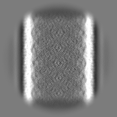
| Title | Cryo-EM of Hyper2 tube, ~24 nm diameter | |||||||||
 Map data Map data | ||||||||||
 Sample Sample |
| |||||||||
 Keywords Keywords | Hyper2 / Protein tube / Helical tube / Strand donation / PROTEIN FIBRIL | |||||||||
| Function / homology | DUF1102 domain-containing protein Function and homology information Function and homology information | |||||||||
| Biological species |   Hyperthermus sp. (archaea) Hyperthermus sp. (archaea) | |||||||||
| Method | helical reconstruction / cryo EM / Resolution: 3.4 Å | |||||||||
 Authors Authors | Sonani RR / Miller JG / Conticello V / Egelman EH | |||||||||
| Funding support |  United States, 1 items United States, 1 items
| |||||||||
 Citation Citation | Journal: Nat Commun / Year: 2025 Title: Donor strand complementation and calcium ion coordination drive the chaperone-free polymerization of archaeal cannulae. Authors: Mike Sleutel / Ravi R Sonani / Jessalyn G Miller / Fengbin Wang / Andres Gonzalez Socorro / Yang Chen / Reece Martin / Borries Demeler / Michael J Rudolph / Vikram Alva / Han Remaut / Edward ...Authors: Mike Sleutel / Ravi R Sonani / Jessalyn G Miller / Fengbin Wang / Andres Gonzalez Socorro / Yang Chen / Reece Martin / Borries Demeler / Michael J Rudolph / Vikram Alva / Han Remaut / Edward H Egelman / Vincent P Conticello /    Abstract: Cannulae are structurally rigid tubular protein filaments that accumulate on the extracellular surface of archaea within the family Pyrodictiaceae during cell growth. These obligate anaerobes ...Cannulae are structurally rigid tubular protein filaments that accumulate on the extracellular surface of archaea within the family Pyrodictiaceae during cell growth. These obligate anaerobes propagate under hyperthermophilic conditions in which cannulae form a biomatrix that interconnects and sustains cells. The persistence of cannulae in this environment suggests that these filaments display significant thermostability, which has attracted technological interest in their development as synthetic protein-based biomaterials. Here, we report cryoEM structural analyses of ex vivo and in vitro assembled recombinant cannulae. We demonstrate that the interactions between protomers in native and recombinant cannulae is based on donor strand complementation (DSC), a form of non-covalent polymerization previously observed for bacterial chaperone-usher pili. Unexpectedly, calcium ion coordination at the subunit interfaces reinforces the network of donor strand interactions in the cannulae. This study provides insight into the mechanism of assembly of cannulae and the structural origin of their high stability and rigidity. | |||||||||
| History |
|
- Structure visualization
| Supplemental images |
|---|

- Downloads & links
-EMDB archive
| Map data | emd_44404.map.gz | 203.8 MB | EMDB map data format | |
|---|---|---|---|---|
| Header (meta data) | emd-44404-v30.xmlemd-44404.xml | 20.1 KB 20.1 KB | Display Display | EMDB header |
| FSC (resolution estimation) | emd_44404_fsc.xml | 12.7 KB | Display | FSC data file |
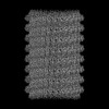
| Images |  emd_44404.png emd_44404.png | 109.1 KB | ||
| Filedesc metadata | emd-44404.cif.gz | 5.9 KB | ||
| Others | emd_44404_additional_1.map.gzemd_44404_half_map_1.map.gzemd_44404_half_map_2.map.gz | 182.7 MB 200.6 MB 200.6 MB | ||
| Archive directory |  http://ftp.pdbj.org/pub/emdb/structures/EMD-44404ftp://ftp.pdbj.org/pub/emdb/structures/EMD-44404 http://ftp.pdbj.org/pub/emdb/structures/EMD-44404ftp://ftp.pdbj.org/pub/emdb/structures/EMD-44404 | HTTPS FTP |
-Related structure data
| Related structure data |  9bacMC  7uiiC  9babC  9dloC  9h8bC C: citing same article ( M: atomic model generated by this map |
|---|---|
| Similar structure data |


-Links
| EMDB pages | EMDB (EBI/PDBe) / EMDataResource |
|---|
-Map
| File | Download / File: emd_44404.map.gz / Format: CCP4 / Size: 216 MB / Type: IMAGE STORED AS FLOATING POINT NUMBER (4 BYTES) | ||||||||||||||||||||||||||||||||||||
|---|---|---|---|---|---|---|---|---|---|---|---|---|---|---|---|---|---|---|---|---|---|---|---|---|---|---|---|---|---|---|---|---|---|---|---|---|---|
| Projections & slices | Image control
Images are generated by Spider. | ||||||||||||||||||||||||||||||||||||
| Voxel size | X=Y=Z: 1.08 Å | ||||||||||||||||||||||||||||||||||||
| Density |
| ||||||||||||||||||||||||||||||||||||
| Symmetry | Space group: 1 | ||||||||||||||||||||||||||||||||||||
| Details | EMDB XML:
|
 Z (Sec.)
 Y (Row.)
 X (Col.)


-Supplemental data
-Additional map: Sharpened Map
| File | emd_44404_additional_1.map | ||||||||||||
|---|---|---|---|---|---|---|---|---|---|---|---|---|---|
| Annotation | Sharpened Map | ||||||||||||
| Projections & Slices |
| ||||||||||||
| Density Histograms |


-Half map: #1
| File | emd_44404_half_map_1.map | ||||||||||||
|---|---|---|---|---|---|---|---|---|---|---|---|---|---|
| Projections & Slices |
| ||||||||||||
| Density Histograms |


-Half map: #2
| File | emd_44404_half_map_2.map | ||||||||||||
|---|---|---|---|---|---|---|---|---|---|---|---|---|---|
| Projections & Slices |
| ||||||||||||
| Density Histograms |


- Sample components
-Entire : Hyper2 tube
| Entire | Name: Hyper2 tube |
|---|---|
| Components |
|
-Supramolecule #1: Hyper2 tube
| Supramolecule | Name: Hyper2 tube / type: complex / ID: 1 / Parent: 0 / Macromolecule list: #1 |
|---|---|
| Source (natural) | Organism: Hyperthermus sp. (archaea) |
-Macromolecule #1: DUF1102 domain-containing protein
| Macromolecule | Name: DUF1102 domain-containing protein / type: protein_or_peptide / ID: 1 / Number of copies: 1 / Enantiomer: LEVO |
|---|---|
| Source (natural) | Organism: Hyperthermus sp. (archaea) |
| Molecular weight | Theoretical: 16.234279 KDa |
| Recombinant expression | Organism:  |
| Sequence | String: MSVTSDRTYY GYGDVSVKNE PVNVVVSPFS FPGANASLSS GGQGVFKKPD WIRVVNQSDV ENVKLEIDWV NANQAANYFD YARILVTGP NGQVKGYLSL QHGKAWITLD AEELREGAVL GAVMYYEVKE GVLASRLPLV FKVRVVETG UniProtKB: DUF1102 domain-containing protein |
-Macromolecule #2: CALCIUM ION
| Macromolecule | Name: CALCIUM ION / type: ligand / ID: 2 / Number of copies: 3 / Formula: CA |
|---|---|
| Molecular weight | Theoretical: 40.078 Da |
-Experimental details
-Structure determination
| Method | cryo EM |
|---|---|
 Processing Processing | helical reconstruction |
| Aggregation state | filament |
-Sample preparation
| Buffer | pH: 8 |
|---|---|
| Vitrification | Cryogen name: ETHANE |
- Electron microscopy
| Microscope | TFS KRIOS |
|---|---|
| Image recording | Film or detector model: GATAN K3 (6k x 4k) / Average electron dose: 50.0 e/Å2 |
| Electron beam | Acceleration voltage: 300 kV / Electron source:  FIELD EMISSION GUN FIELD EMISSION GUN |
| Electron optics | Illumination mode: FLOOD BEAM / Imaging mode: BRIGHT FIELD / Nominal defocus max: 2.2 µm / Nominal defocus min: 1.2 µm |
| Experimental equipment |  Model: Titan Krios / Image courtesy: FEI Company |

