 ムービー
ムービー コントローラー
 構造ビューア
 EMN検索について


-検索条件
-検索結果
検索 (著者・登録者: lewis & t)の結果200件中、1から50件目までを表示しています

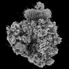
EMDB-50358: 
In vitro-induced genome-releasing intermediate of Rhodobacter microvirus Ebor computed with C5 symmetry
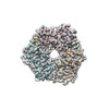
EMDB-19568:
DtpB hexamer from Streptomyces lividans
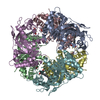
PDB-8rwy:
DtpB hexamer from Streptomyces lividans
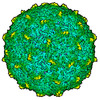
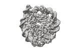
EMDB-50356:
Empty capsid of Rhodobacter microvirus Ebor computed with I4 symmetry
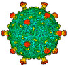
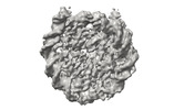
EMDB-50357:
Native capsid of Rhodobacter microvirus Ebor computed with I4 symmetry

EMDB-50359:
Rhodobacter microvirus Ebor attached to B10 host cell reconstructed by single particle analysis with applied C5 symmetry

EMDB-50360:
Rhodobacter microvirus Ebor attached to the outer membrane vesicle

EMDB-50361:
Rhodobacter microvirus Ebor attached to the host cell reconstructed by subtomogram averaging

PDB-9ffg:
Empty capsid of Rhodobacter microvirus Ebor computed with I4 symmetry

PDB-9ffh:
Native capsid of Rhodobacter microvirus Ebor computed with I4 symmetry
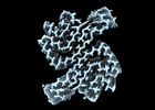
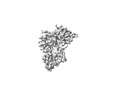
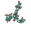
EMDB-19986:
Structure of recombinant alpha-synuclein fibrils 1B capable of seeding GCIs in vivo

PDB-9euu:
Structure of recombinant alpha-synuclein fibrils 1B capable of seeding GCIs in vivo

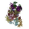
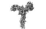
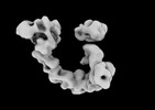
EMDB-17449:
S. cerevisiae nexus-sCMGE after DNA replication initiation

EMDB-17458:
S. cerevisiae ssDNA-sCMGE after DNA replication initiation

EMDB-17459:
S. cerevisiae consensus-sCMGE on ssDNA after DNA replication initiation

EMDB-17460:
S. cerevisiae sCMGE with N-ter Mcm10 density

PDB-8p5e:
S. cerevisiae nexus-sCMGE after DNA replication initiation
PDB-8p62:
S. cerevisiae ssDNA-sCMGE after DNA replication initiation

PDB-8p63:
S. cerevisiae consensus-sCMGE on ssDNA after DNA replication initiation

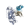
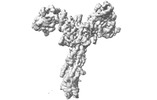
EMDB-40825:
10E8-GT10.2 immunogen in complex with human Fab 10E8 and mouse Fab W6-10
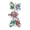
PDB-8sx3:
10E8-GT10.2 immunogen in complex with human Fab 10E8 and mouse Fab W6-10

PDB-8qox:
Two-component assembly of SlaA and SlaB S-layer proteins of Sulfolobus acidocaldarius

PDB-8qp0:
A hexamer pore in the S-layer of Sulfolobus acidocaldarius formed by SlaA protein
EMDB-18127:
S-layer of archaeon Sulfolobus acidocaldarius by subtomogram averaging
EMDB-19199:
Structure of the rabbit 80S ribosome stalled on a 2-TMD rhodopsin intermediate in complex with Sec61, bound TRAP-alpha TMD
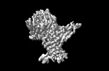
EMDB-41810:
Cryo-EM structure of vaccine-elicited CD4 binding site antibody DH1285 bound to HIV-1 CH505TFchim.6R.SOSIP.664v4.1 Env Local Refinement

EMDB-41820:
Cryo-EM structure of vaccine-elicited CD4 binding site antibody DH1285 bound to HIV-1 CH505TFchim.6R.SOSIP.664v4.1 Env

EMDB-41823:
Cryo-EM structure of vaccine-elicited CD4 binding site antibody DH1285 bound to HIV-1 CH505TFchim.6R.SOSIP.664v4.1 Env

EMDB-41838:
Cryo-EM structure of vaccine-elicited CD4 binding site antibody DH1285 bound to partially open HIV-1 CH505TFchim.6R.SOSIP.664v4.1 Env
PDB-8u1d:
Cryo-EM structure of vaccine-elicited CD4 binding site antibody DH1285 bound to HIV-1 CH505TFchim.6R.SOSIP.664v4.1 Env Local Refinement
EMDB-15530:
S-layer protein SlaA from Sulfolobus acidocaldarius at pH 10.0
EMDB-15531:
S-layer protein SlaA from Sulfolobus acidocaldarius at pH 7.0

PDB-8an2:
S-layer protein SlaA from Sulfolobus acidocaldarius at pH 10.0

PDB-8an3:
S-layer protein SlaA from Sulfolobus acidocaldarius at pH 7.0
EMDB-27703:
Structure of RBD directed antibody DH1047 in complex with SARS-CoV-2 spike: Local refinement of RBD-Fab interace
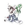
PDB-8dtk:
Structure of RBD directed antibody DH1047 in complex with SARS-CoV-2 spike: Local refinement of RBD-Fab interace

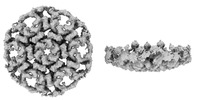
EMDB-41182:
Cryo-EM map of the Unmodified nucleosome core particle in 100 mM KCl with local resolution values
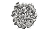
EMDB-41183:
Cryo-EM map of the PARylated nucleosome core particle in 100 mM KCl with local resolution values
EMDB-41184:
Cryo-EM map of the Unmodified nucleosome core particle in 5 mM KCl with local resolution values
EMDB-41178:
Cryo-EM map of the PARylated nucleosome core particle in 5 mM KCl with local resolution values

EMDB-16519:
Slipper limpet hemocyanin didecamer

EMDB-16523:
Slipper limpet hemocyanin tridecamer

EMDB-14635:
S-layer protein SlaA from Sulfolobus acidocaldarius at pH 4.0
PDB-7zcx:
S-layer protein SlaA from Sulfolobus acidocaldarius at pH 4.0
EMDB-29101:
EGFR:Degrader:VHL:Elongin-B/C:Cul2

EMDB-28754:
Client-bound structure of a DegP trimer within a 12mer cage

EMDB-28781:
Structure of a 12mer DegP cage bound to the client protein hTRF1

EMDB-28800:
Structure of an 18mer DegP cage bound to the client protein hTRF1

EMDB-28801:
Structure of a 24mer DegP cage bound to the client protein hTRF1

EMDB-28806:
Structure of a 30mer DegP cage bound to the client protein hTRF1
ページ:
 wwPDBはEMDBデータモデルのバージョン3へ移行します
