Journal: Protein Sci / Year: 2024 Title: The oligomeric states of dye-decolorizing peroxidases from Streptomyces lividans and their implications for mechanism of substrate oxidation. Authors: Marina Lučić / Thomas Allport / Thomas A Clarke / Lewis J Williams / Michael T Wilson / Amanda K Chaplin / Jonathan A R Worrall / Abstract: A common evolutionary mechanism in biology to drive function is protein oligomerization. In prokaryotes, the symmetrical assembly of repeating protein units to form homomers is widespread, yet ...A common evolutionary mechanism in biology to drive function is protein oligomerization. In prokaryotes, the symmetrical assembly of repeating protein units to form homomers is widespread, yet consideration in vitro of whether such assemblies have functional or mechanistic consequences is often overlooked. Dye-decolorizing peroxidases (DyPs) are one such example, where their dimeric α + β barrel units can form various oligomeric states, but the oligomer influence, if any, on mechanism and function has received little attention. In this work, we have explored the oligomeric state of three DyPs found in Streptomyces lividans, each with very different mechanistic behaviors in their reactions with hydrogen peroxide and organic substrates. Using analytical ultracentrifugation, we reveal that except for one of the A-type DyPs where only a single sedimenting species is detected, oligomer states ranging from homodimers to dodecamers are prevalent in solution. Using cryo-EM on preparations of the B-type DyP, we determined a 3.02 Å resolution structure of a hexamer assembly that corresponds to the dominant oligomeric state in solution as determined by analytical ultracentrifugation. Furthermore, cryo-EM data detected sub-populations of higher-order oligomers, with one of these formed by an arrangement of two B-type DyP hexamers to give a dodecamer assembly. Our solution and structural insights of these oligomer states provide a new framework to consider previous mechanistic studies of these DyP members and are discussed in terms of long-range electron transfer for substrate oxidation and in the "storage" of oxidizable equivalents on the heme until a two-electron donor is available.
A: Dyp-type peroxidase family B: Dyp-type peroxidase family C: Dyp-type peroxidase family D: Dyp-type peroxidase family E: Dyp-type peroxidase family F: Dyp-type peroxidase family hetero molecules
In the structure databanks used in Yorodumi, some data are registered as the other names, "COVID-19 virus" and "2019-nCoV". Here are the details of the virus and the list of structure data.
Jan 31, 2019. EMDB accession codes are about to change! (news from PDBe EMDB page)
EMDB accession codes are about to change! (news from PDBe EMDB page)
The allocation of 4 digits for EMDB accession codes will soon come to an end. Whilst these codes will remain in use, new EMDB accession codes will include an additional digit and will expand incrementally as the available range of codes is exhausted. The current 4-digit format prefixed with “EMD-” (i.e. EMD-XXXX) will advance to a 5-digit format (i.e. EMD-XXXXX), and so on. It is currently estimated that the 4-digit codes will be depleted around Spring 2019, at which point the 5-digit format will come into force.
The EM Navigator/Yorodumi systems omit the EMD- prefix.
Related info.:Q: What is EMD? / ID/Accession-code notation in Yorodumi/EM Navigator
Yorodumi is a browser for structure data from EMDB, PDB, SASBDB, etc.
This page is also the successor to EM Navigator detail page, and also detail information page/front-end page for Omokage search.
The word "yorodu" (or yorozu) is an old Japanese word meaning "ten thousand". "mi" (miru) is to see.
Related info.:EMDB / PDB / SASBDB / Comparison of 3 databanks / Yorodumi Search / Aug 31, 2016. New EM Navigator & Yorodumi / Yorodumi Papers / Jmol/JSmol / Function and homology information / Changes in new EM Navigator and Yorodumi
 Movie
Movie Controller


 Open data
 Basic information
 Components
 Keywords
 Function and homology information
 Streptomyces lividans (bacteria)
 Authors
 Citation
 Structure visualization
 Downloads & links
 Other downloads
 PDBj
PDBj Assembly
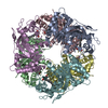

 Mass: 616.487 Da / Num. of mol.: 6 / Source method: obtained synthetically / Formula: C34H32FeN4O4 / Feature type: SUBJECT OF INVESTIGATION
 Sample preparation
 Electron microscopy imaging
 FIELD EMISSION GUN / Accelerating voltage: 300 kV / Illumination mode: FLOOD BEAM
 Processing