Movie
Movie Controller


+ Open data

- Basic information
| Entry |  | |||||||||||||||
|---|---|---|---|---|---|---|---|---|---|---|---|---|---|---|---|---|
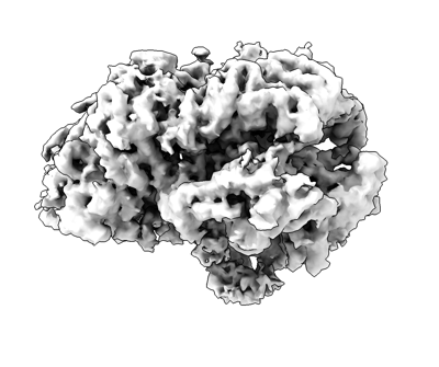
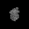
| Title | S. cerevisiae sCMGE with N-ter Mcm10 density | |||||||||||||||
 Map data Map data | EM map of sCMG10 | |||||||||||||||
 Sample Sample |
| |||||||||||||||
 Keywords Keywords | Saccharomyces cerevisiae / helicase / CMGE / initiation of DNA replication / DNA / DNA unwinding / REPLICATION / Mcm10 | |||||||||||||||
| Biological species |  | |||||||||||||||
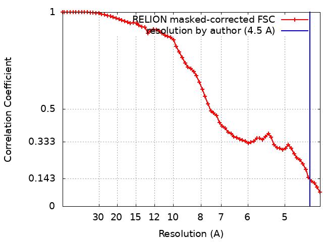
| Method | single particle reconstruction / cryo EM / Resolution: 4.5 Å | |||||||||||||||
 Authors Authors | Henrikus SS / Willhoft O | |||||||||||||||
| Funding support |  United Kingdom, European Union, United Kingdom, European Union,  France, 4 items France, 4 items
| |||||||||||||||
 Citation Citation | Journal: Nat Struct Mol Biol / Year: 2024 Title: Unwinding of a eukaryotic origin of replication visualized by cryo-EM. Authors: Sarah S Henrikus / Marta H Gross / Oliver Willhoft / Thomas Pühringer / Jacob S Lewis / Allison W McClure / Julia F Greiwe / Giacomo Palm / Andrea Nans / John F X Diffley / Alessandro Costa /   Abstract: To prevent detrimental chromosome re-replication, DNA loading of a double hexamer of the minichromosome maintenance (MCM) replicative helicase is temporally separated from DNA unwinding. Upon S-phase ...To prevent detrimental chromosome re-replication, DNA loading of a double hexamer of the minichromosome maintenance (MCM) replicative helicase is temporally separated from DNA unwinding. Upon S-phase transition in yeast, DNA unwinding is achieved in two steps: limited opening of the double helix and topological separation of the two DNA strands. First, Cdc45, GINS and Polε engage MCM to assemble a double CMGE with two partially separated hexamers that nucleate DNA melting. In the second step, triggered by Mcm10, two CMGEs separate completely, eject the lagging-strand template and cross paths. To understand Mcm10 during helicase activation, we used biochemical reconstitution with cryogenic electron microscopy. We found that Mcm10 splits the double CMGE by engaging the N-terminal homo-dimerization face of MCM. To eject the lagging strand, DNA unwinding is started from the N-terminal side of MCM while the hexamer channel becomes too narrow to harbor duplex DNA. | |||||||||||||||
| History |
|
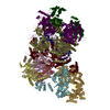
- Structure visualization
| Supplemental images |
|---|
- Downloads & links
-EMDB archive
| Map data | emd_17460.map.gz | 2.1 MB |  EMDB map data format EMDB map data format | |
|---|---|---|---|---|
| Header (meta data) | emd-17460-v30.xmlemd-17460.xml | 34 KB 34 KB | Display Display | EMDB header |
| FSC (resolution estimation) | emd_17460_fsc.xml | 6.4 KB | Display | FSC data file |
| Images |  emd_17460.png emd_17460.png | 82.4 KB | ||
| Filedesc metadata | emd-17460.cif.gz | 9.3 KB | ||
| Others | emd_17460_half_map_1.map.gzemd_17460_half_map_2.map.gz | 17 MB 17 MB | ||
| Archive directory |  http://ftp.pdbj.org/pub/emdb/structures/EMD-17460ftp://ftp.pdbj.org/pub/emdb/structures/EMD-17460 http://ftp.pdbj.org/pub/emdb/structures/EMD-17460ftp://ftp.pdbj.org/pub/emdb/structures/EMD-17460 | HTTPS FTP |
-Related structure data


-Links
| EMDB pages | EMDB (EBI/PDBe) / EMDataResource |
|---|---|
| Related items in Molecule of the Month |


-Map
| File | Download / File: emd_17460.map.gz / Format: CCP4 / Size: 22.2 MB / Type: IMAGE STORED AS FLOATING POINT NUMBER (4 BYTES) | ||||||||||||||||||||||||||||||||||||
|---|---|---|---|---|---|---|---|---|---|---|---|---|---|---|---|---|---|---|---|---|---|---|---|---|---|---|---|---|---|---|---|---|---|---|---|---|---|
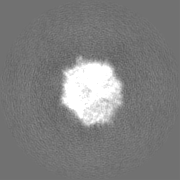
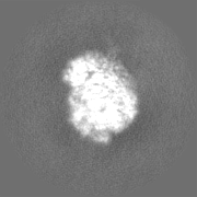
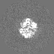
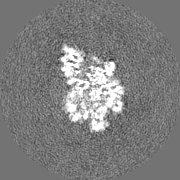
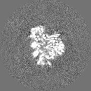
| Annotation | EM map of sCMG10 | ||||||||||||||||||||||||||||||||||||
| Projections & slices | Image control
Images are generated by Spider. | ||||||||||||||||||||||||||||||||||||
| Voxel size | X=Y=Z: 2.16 Å | ||||||||||||||||||||||||||||||||||||
| Density |
| ||||||||||||||||||||||||||||||||||||
| Symmetry | Space group: 1 | ||||||||||||||||||||||||||||||||||||
| Details | EMDB XML:
|
 Z (Sec.)
 Y (Row.)
 X (Col.)


-Supplemental data
-Half map: EM half map 2 of sCMG10
| File | emd_17460_half_map_1.map | ||||||||||||
|---|---|---|---|---|---|---|---|---|---|---|---|---|---|
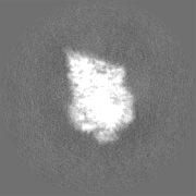
| Annotation | EM half map 2 of sCMG10 | ||||||||||||
| Projections & Slices |
| ||||||||||||
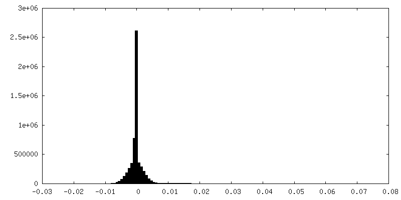
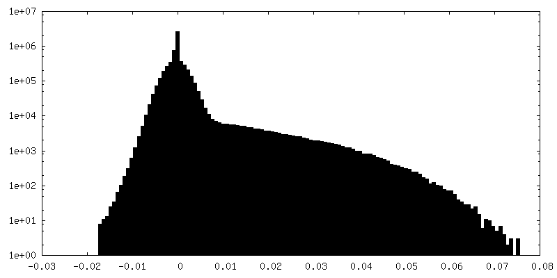
| Density Histograms |
- Sample components
+Entire : Complex of Mcm2-7, Cdc45, GINS (Psf1-3, Sld5) DNA polymerase epsi...
+Supramolecule #1: Complex of Mcm2-7, Cdc45, GINS (Psf1-3, Sld5) DNA polymerase epsi...
+Supramolecule #2: DNA replication complex GINS containing Psf1, Psf2, Psf3 and Sld5
+Supramolecule #3: DNA replication licensing factors MCM2-7
+Supramolecule #4: DNA polymerase epsilon contains catalytic subunit A and non-catal...
+Supramolecule #5: DNA
+Supramolecule #6: Cdc45
+Macromolecule #1: DNA replication licensing factor MCM2

+Macromolecule #2: DNA replication licensing factor MCM3
+Macromolecule #3: DNA replication licensing factor MCM4
+Macromolecule #4: DNA replication licensing factor MCM5
+Macromolecule #5: DNA replication licensing factor MCM6
+Macromolecule #6: Cell division control protein 45
+Macromolecule #7: DNA replication complex GINS protein PSF1
+Macromolecule #8: DNA replication complex GINS protein PSF2
+Macromolecule #9: DNA replication complex GINS protein PSF3
+Macromolecule #10: DNA replication complex GINS protein SLD5
+Macromolecule #11: Minichromosome maintenance protein 10
+Macromolecule #14: DNA replication licensing factor MCM7
+Macromolecule #12: DNA strand 1
+Macromolecule #13: DNA strand 2
-Experimental details
-Structure determination
| Method | cryo EM |
|---|---|
 Processing Processing | single particle reconstruction |
| Aggregation state | particle |
-Sample preparation
| Buffer | pH: 7.6 |
|---|---|
| Vitrification | Cryogen name: ETHANE / Instrument: FEI VITROBOT MARK IV |
- Electron microscopy
| Microscope | FEI TITAN KRIOS |
|---|---|
| Image recording | Film or detector model: GATAN K2 SUMMIT (4k x 4k) / Average electron dose: 1.67 e/Å2 |
| Electron beam | Acceleration voltage: 300 kV / Electron source:  FIELD EMISSION GUN FIELD EMISSION GUN |
| Electron optics | Illumination mode: SPOT SCAN / Imaging mode: BRIGHT FIELD / Nominal defocus max: 3.3000000000000003 µm / Nominal defocus min: 1.8 µm |
| Experimental equipment |  Model: Titan Krios / Image courtesy: FEI Company |