 Movie
Movie Controller
 Structure viewers
 About EMN search


-Search query
-Search result
Showing 1 - 50 of 536 items for (author: lak & p)
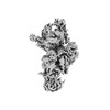
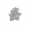
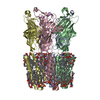
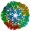
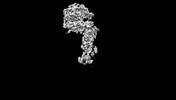
EMDB-18438: 
mt-SSU assembly intermediate in GTPBP8 knock-out cells, state 1
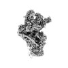
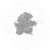
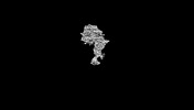
EMDB-18439:
mt-SSU assembly intermediate in GTPBP8 knock-out cells, state 2
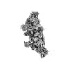
EMDB-18440:
mt-SSU assembly intermediate in GTPBP8 knock-out cells, state 3
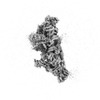
EMDB-18443:
mt-SSU assembly intermediate in GTPBP8 knock-out cells, state 4
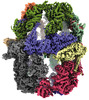
EMDB-18460:
mt-LSU assembly intermediate in GTPBP8 knock-out cells, state 1
EMDB-18461:
mt-LSU assembly intermediate in GTPBP8 knock-out cells, state 2
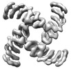
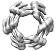
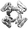
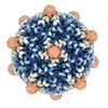
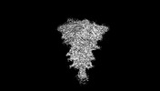
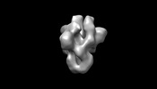
EMDB-28966:
CryoEM map of de novo designed oligomeric protein C4-71_6x
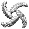
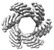
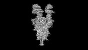
EMDB-28967:
CryoEM map of de novo designed oligomeric protein C4-71_8x
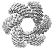
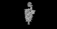
EMDB-28968:
CryoEM map of de novo designed oligomeric protein C6-71
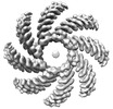
EMDB-28969:
CryoEM map of de novo designed oligomeric protein C6-71_6x
EMDB-28970:
CryoEM map of de novo designed oligomeric protein C6-71_8x

EMDB-28971:
CryoEM map of de novo designed oligomeric protein C8-71_6x
EMDB-28972:
CryoEM map of de novo designed oligomeric protein C8-71_8x
EMDB-28973:
CryoEM map of de novo designed oligomeric protein C4-81

EMDB-28974:
CryoEM map of designed oligomeric protein C4-71

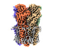
EMDB-35163:
Cryo-EM structure of nanodisc (PE:PS:PC) reconstituted GLIC at pH 5.5

EMDB-35164:
Cryo-EM structure of nanodisc (PE:PS:PC) reconstituted GLIC at pH 4 in closed state
EMDB-36339:
Cryo-EM structure of nanodisc (PE:PS:PC) reconstituted GLIC at pH 2.5
EMDB-37446:
Cryo-EM structure of nanodisc (PE:PS:PC) reconstituted GLIC at pH 4 in intermediate state
EMDB-37447:
Cryo-EM structure of nanodisc (PE:PS:PC) reconstituted GLIC at pH 4 in open state
PDB-8i47:
Cryo-EM structure of nanodisc (PE:PS:PC) reconstituted GLIC at pH 5.5
PDB-8i48:
Cryo-EM structure of nanodisc (PE:PS:PC) reconstituted GLIC at pH 4 in closed state
PDB-8jj3:
Cryo-EM structure of nanodisc (PE:PS:PC) reconstituted GLIC at pH 2.5
PDB-8wcq:
Cryo-EM structure of nanodisc (PE:PS:PC) reconstituted GLIC at pH 4 in intermediate state
PDB-8wcr:
Cryo-EM structure of nanodisc (PE:PS:PC) reconstituted GLIC at pH 4 in open state

EMDB-35161:
Cryo-EM structure of nanodisc (asolectin) reconstituted GLIC at pH 7.5

EMDB-35162:
Cryo-EM structure of nanodisc (PE:PS:PC) reconstituted GLIC at pH 7.5

PDB-8i41:
Cryo-EM structure of nanodisc (asolectin) reconstituted GLIC at pH 7.5

PDB-8i42:
Cryo-EM structure of nanodisc (PE:PS:PC) reconstituted GLIC at pH 7.5

EMDB-18659:
Cryo-EM Structure of Human Kv3.1 in Complex with Modulator AUT1

EMDB-18660:
Cryo-EM Structure of Human Kv3.1 in Complex with Modulator AUT5

PDB-8quc:
Cryo-EM Structure of Human Kv3.1 in Complex with Modulator AUT1

PDB-8qud:
Cryo-EM Structure of Human Kv3.1 in Complex with Modulator AUT5
EMDB-19477:
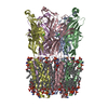
Saccharomyces cerevisiae FAS type I
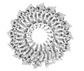
EMDB-19489:
Tobacco mosaic virus from scanning transmission electron microscopy at CSA=2.0 mrad
EMDB-42812:
PNMA2 capsid, overall icosahedral map

EMDB-42815:
PNMA2 capsid, focussed refinement of a pentamer (C5 symmetry)
PDB-8uyo:
Structure of a recombinant human PNMA2 capsid

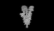
EMDB-16916:
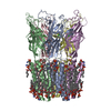
Bipartite interaction of TOPBP1 with the GINS complex

PDB-8ok2:
Bipartite interaction of TOPBP1 with the GINS complex
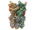
EMDB-43320:
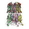
Cryo-EM structure of SARS-CoV-2 XBB.1.5 spike protein
EMDB-43321:
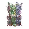
Cryo-EM structure of SARS-CoV-2 XBB.1.5 spike protein in complex with mouse ACE2 (conformation 2)
EMDB-43322:
Cryo-EM structure of SARS-CoV-2 XBB.1.5 spike protein in complex with mouse ACE2 (conformation 1)
EMDB-43323:
Cryo-EM structure of SARS-CoV-2 XBB.1.5 spike protein in complex with mouse ACE2 (focused refinement of RBD and mouse ACE2)
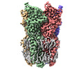
EMDB-43324:
Cryo-EM structure of SARS-CoV-2 XBB.1.5 spike protein in complex with human ACE2
EMDB-43325:
Cryo-EM structure of SARS-CoV-2 XBB.1.5 spike protein in complex with human ACE2 (focused refinement of RBD and ACE2)
EMDB-43326:
Negative Stain EM Reconstructions of SARS-CoV-2 spike proteins mixed with polyclonal antibodies from donor 4.

PDB-8vkk:
Cryo-EM structure of SARS-CoV-2 XBB.1.5 spike protein
PDB-8vkl:
Cryo-EM structure of SARS-CoV-2 XBB.1.5 spike protein in complex with mouse ACE2 (conformation 2)

PDB-8vkm:
Cryo-EM structure of SARS-CoV-2 XBB.1.5 spike protein in complex with mouse ACE2 (conformation 1)
Pages:
 wwPDB to switch to version 3 of the EMDB data model
