 ムービー
ムービー コントローラー
 構造ビューア
 EMN検索について


-検索条件
-検索結果
検索 (著者・登録者: fujii & a)の結果全42件を表示しています
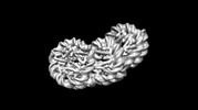
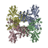
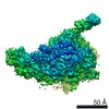
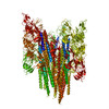
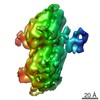
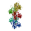
EMDB-35448: 
Overlapping tri-nucleosome
PDB-8ihl:
Overlapping tri-nucleosome
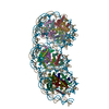
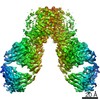
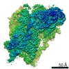
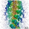
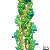
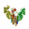
EMDB-32588:
The Cryo-EM structure of siphonaxanthin chlorophyll a/b type light-harvesting complex II
PDB-7wlm:
The Cryo-EM structure of siphonaxanthin chlorophyll a/b type light-harvesting complex II
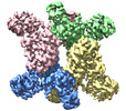
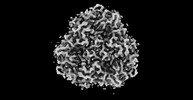
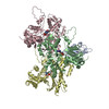
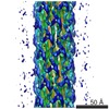
EMDB-32381:
Trichodesmium erythraeum cyanophycin synthetase 1 (TeCphA1)
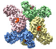
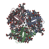
EMDB-32382:
Trichodesmium erythraeum cyanophycin synthetase 1 (TeCphA1) with ATPgammaS
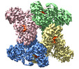
EMDB-32383:
Trichodesmium erythraeum cyanophycin synthetase 1 (TeCphA1) with ATPgammaS, 4x(beta-Asp-Arg), and aspartate
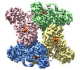
EMDB-32384:
Trichodesmium erythraeum cyanophycin synthetase 1 (TeCphA1) with ATPgammaS and 4x(beta-Asp-Arg)
PDB-7wac:
Trichodesmium erythraeum cyanophycin synthetase 1 (TeCphA1)
PDB-7wad:
Trichodesmium erythraeum cyanophycin synthetase 1 (TeCphA1) with ATPgammaS

PDB-7wae:
Trichodesmium erythraeum cyanophycin synthetase 1 (TeCphA1) with ATPgammaS, 4x(beta-Asp-Arg), and aspartate
PDB-7waf:
Trichodesmium erythraeum cyanophycin synthetase 1 (TeCphA1) with ATPgammaS and 4x(beta-Asp-Arg)
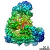
EMDB-0869:
Cryo-EM structure of the MgtE Mg2+ channel under Mg2+-free conditions

PDB-6lbh:
Cryo-EM structure of the MgtE Mg2+ channel under Mg2+-free conditions
EMDB-11562:
Mouse Hoxa9 IRES like element bound to the human 80S ribosome
EMDB-11563:
Mouse Hoxa9 IRES-like element bound to the human 40S ribosomal subunit head - IRES binding site
EMDB-11564:
Mouse Hoxa9 IRES-like element bound to the human 40S ribosomal subunit - 40S head
EMDB-11565:
Mouse Hoxa9 IRES-like element bound to the human 40S ribosomal subunit - 40S body
EMDB-11566:
Hoxa9 IRES P4 stem-loop bound to the 40S ribosomal subunit - 40S head
EMDB-11567:
Hoxa9 IRES P4 stem-loop bound to the 40S ribosomal subunit - 40S body

EMDB-11568:
human 40S ribosomal subunit (control)

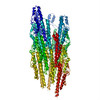
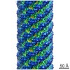
EMDB-0728:
Structure of human cardiac thin filament in the calcium free state
EMDB-0729:
Structure of human cardiac thin filament in the calcium bound state

PDB-6kn7:
Structure of human cardiac thin filament in the calcium free state

PDB-6kn8:
Structure of human cardiac thin filament in the calcium bound state
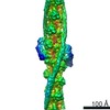
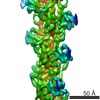
EMDB-6683:
The 7 angstrom resolution CryoEM map of the bacterial flagellar polyrod
PDB-5wrh:
FlgG structure based on the CryoEM map of the bacterial flagellar polyrod
EMDB-6664:
The structure of rabbit skeletal muscle actomyosin rigor complex at 5.2 angstrom.

PDB-5h53:
The structure of rabbit skeletal muscle actomyosin rigor complex at 5.2 angstrom.

EMDB-6102:
Electron cryo-microscopy of DNGR-1 in complex with F-actin
PDB-3j82:
Electron cryo-microscopy of DNGR-1 in complex with F-actin
EMDB-2929:
Mechanism of Ubiquitin Ligation to a Substrate by human Anaphase-Promoting Complex
EMDB-5931:
Dictyostelium dynein microtubule binding domain bound to a 15-protofilament microtubule

PDB-3j6p:
Pseudo-atomic model of dynein microtubule binding domain-tubulin complex based on a cryoEM map

PDB-4a6j:
Structural model of ParM filament based on CryoEM map
EMDB-1980:
CryoEM map of ParM filament at 7.2 Angstrom resolution
PDB-3j0r:
Model of a type III secretion system needle based on a 7 Angstrom resolution cryoEM map
EMDB-5352:
Structure of a type III secretion needle
PDB-3mfp:
Atomic model of F-actin based on a 6.6 angstrom resolution cryoEM map
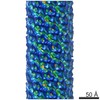
EMDB-5168:
Direct visualization of secondary structures of F-actin by electron cryomicroscopy
PDB-3a69:
Atomic model of the bacterial flagellar hook based on docking an X-ray derived structure and terminal two alpha-helices into an 7.1 angstrom resolution cryoEM map
EMDB-1647:
Specific arrangement of alpha-helical coiled coils in the core domain of the bacterial flagellar hook for the universal joint function
 wwPDBはEMDBデータモデルのバージョン3へ移行します
